RARENET recrute !
Rarenet recrute un assistant ingénieur dans le cadre du projet Genodent.
Pour en savoir plus, télécharger la fiche de poste.
Rarenet recrute un assistant ingénieur dans le cadre du projet Genodent.
Pour en savoir plus, télécharger la fiche de poste.
“Latent-transforming growth factor beta-binding protein 3 (LTBP-3) is important for craniofacial morphogenesis and hard tissue mineralization, as it is essential for activation of transforming growth factor-β (TGF-β). To investigate the role of LTBP-3 in tooth formation we performed micro-computed tomography (micro-CT), histology, and scanning electron microscopy analyses of adult Ltbp3-/- mice. The Ltbp3-/- mutants presented with unique craniofacial malformations and reductions in enamel formation that began at the matrix formation stage. Organization of maturation-stage ameloblasts was severely disrupted. The lateral side of the incisor was affected most. Reduced enamel mineralization, modification of the enamel prism pattern, and enamel nodules were observed throughout the incisors, as revealed by scanning electron microscopy. Molar roots had internal irregular bulbous-like formations. The cementum thickness was reduced, and microscopic dentinal tubules showed minor nanostructural changes. Thus, LTBP-3 is required for ameloblast differentiation and for the formation of decussating enamel prisms, to prevent enamel nodule formation, and for proper root morphogenesis. Also, and consistent with the role of TGF-β signaling during mineralization, almost all craniofacial bone components were affected in Ltbp3-/- mice, especially those involving the upper jaw and snout. This mouse model demonstrates phenotypic overlap with Verloes Bourguignon syndrome, also caused by mutation of LTBP3, which is hallmarked by craniofacial anomalies and amelogenesis imperfecta phenotypes.”
Read the article: https://www.ncbi.nlm.nih.gov/pmc/articles/PMC5260799/
Retrouvez le descriptif du projet en format PDF
“WNT10A gene encodes a canonical wingless pathway signaling molecule involved in cell fate specification as well as morphogenetic patterning of the developing ectoderm, nervous system, skeleton, and tooth. In patients, WNT10A mutations are responsible for ectodermal-derived pathologies including isolated hypo-oligodontia, tricho-odonto-onycho-dermal dysplasia and Schöpf-Schulz-Passarge syndrome (SSPS). Here we describe the dental, ectodermal, and extra-ectodermal phenotypic features of a cohort of 41 patients from 32 unrelated families. Correlations with WNT10A molecular status (heterozygous carrier, compound heterozygous, homozygous) and patient’s phenotypes were performed. Mild to severe oligodontia was observed in all patients bearing biallelic WNT10A mutations. However, patients with compound heterozygous mutations presented no significant difference in phenotypes compared with homozygous individuals. Anomalies in tooth morphology were frequently observed with heterozygous patients displaying hypodontia. No signs of SSPS, especially eyelids cysts, were detected in our cohort. Interestingly, extra-ectodermal signs consisted of skeletal, neurological and vascular anomalies, the latter suggesting a wider phenotypic spectrum associated with WNT10A mutations. Indeed, the Wnt pathway plays a crucial role in skeletal development, lipid metabolism, and neurogenesis, potentially explaining patient’s clinical manifestations.”
“Abnormalities of enamel matrix proteins deposition, mineralization, or degradation during tooth development are responsible for a spectrum of either genetic diseases termed Amelogenesis imperfecta or acquired enamel defects. To assess if environmental/nutritional factors can exacerbate enamel defects, we investigated the role of the active form of vitamin A, retinoic acid (RA). Robust expression of RA-degrading enzymes Cyp26b1 and Cyp26c1 in developing murine teeth suggested RA excess would reduce tooth hard tissue mineralization, adversely affecting enamel. We employed a protocol where RA was supplied to pregnant mice as a food supplement, at a concentration estimated to result in moderate elevations in serum RA levels. This supplementation led to severe enamel defects in adult mice born from pregnant dams, with most severe alterations observed for treatments from embryonic day (E)12.5 to E16.5. We identified the enamel matrix proteins enamelin (Enam), ameloblastin (Ambn), and odontogenic ameloblast-associated protein (Odam) as target genes affected by excess RA, exhibiting mRNA reductions of over 20-fold in lower incisors at E16.5. RA treatments also affected bone formation, reducing mineralization. Accordingly, craniofacial ossification was drastically reduced after 2 days of treatment (E14.5). Massive RNA-sequencing (RNA-seq) was performed on E14.5 and E16.5 lower incisors. Reductions in Runx2 (a key transcriptional regulator of bone and enamel differentiation) and its targets were observed at E14.5 in RA-exposed embryos. RNA-seq analysis further indicated that bone growth factors, extracellular matrix, and calcium homeostasis were perturbed. Genes mutated in human AI (ENAM, AMBN, AMELX, AMTN, KLK4) were reduced in expression at E16.5. Our observations support a model in which elevated RA signaling at fetal stages affects dental cell lineages. Thereafter enamel protein production is impaired, leading to permanent enamel alterations.”
Read the article: https://www.ncbi.nlm.nih.gov/pmc/articles/PMC5217128/
Abstract:
Kohlschütter-Tönz syndrome (KTZS) is a rare autosomal-recessive disease characterised by epileptic encephalopathy, intellectual disability and amelogenesis imperfecta (AI). It is frequently caused by biallelic mutations in ROGDI. Here, we report on individuals with ROGDI-negative KTZS carrying biallelic SLC13A5 mutations.
In the present cohort study, nine individuals from four families with the clinical diagnosis of KTZS and absence of ROGDI mutations as well as one patient with unexplained epileptic encephalopathy were investigated by clinical and dental evaluation, parametric linkage analysis (one family), and exome and/or Sanger sequencing. Dental histological investigations were performed on teeth from individuals with SLC13A5-associated and ROGDI-associated KTZS.
Biallelic mutations in SLC13A5 were identified in 10 affected individuals. Epileptic encephalopathy usually presents in the neonatal and (less frequently) early infantile period. Yellowish to orange discolouration of both deciduous and permanent teeth, as well as wide interdental spaces and abnormal crown forms are major clinical signs of individuals with biallelic SLC13A5 mutations. Histological dental investigations confirmed the clinical diagnosis of hypoplastic AI. In comparison, the histological evaluation of a molar assessed from an individual with ROGDI-associated KTZS revealed hypocalcified AI.
We conclude that SLC13A5 is the second major gene associated with the clinical diagnosis of KTZS, characterised by neonatal epileptic encephalopathy and hypoplastic AI. Careful clinical and dental delineation provides clues whether ROGDI or SLC13A5 is the causative gene. Hypersensitivity of teeth as well as high caries risk requires individual dental prophylaxis and attentive dental management.”
Read the article: http://jmg.bmj.com/content/54/1/54.long
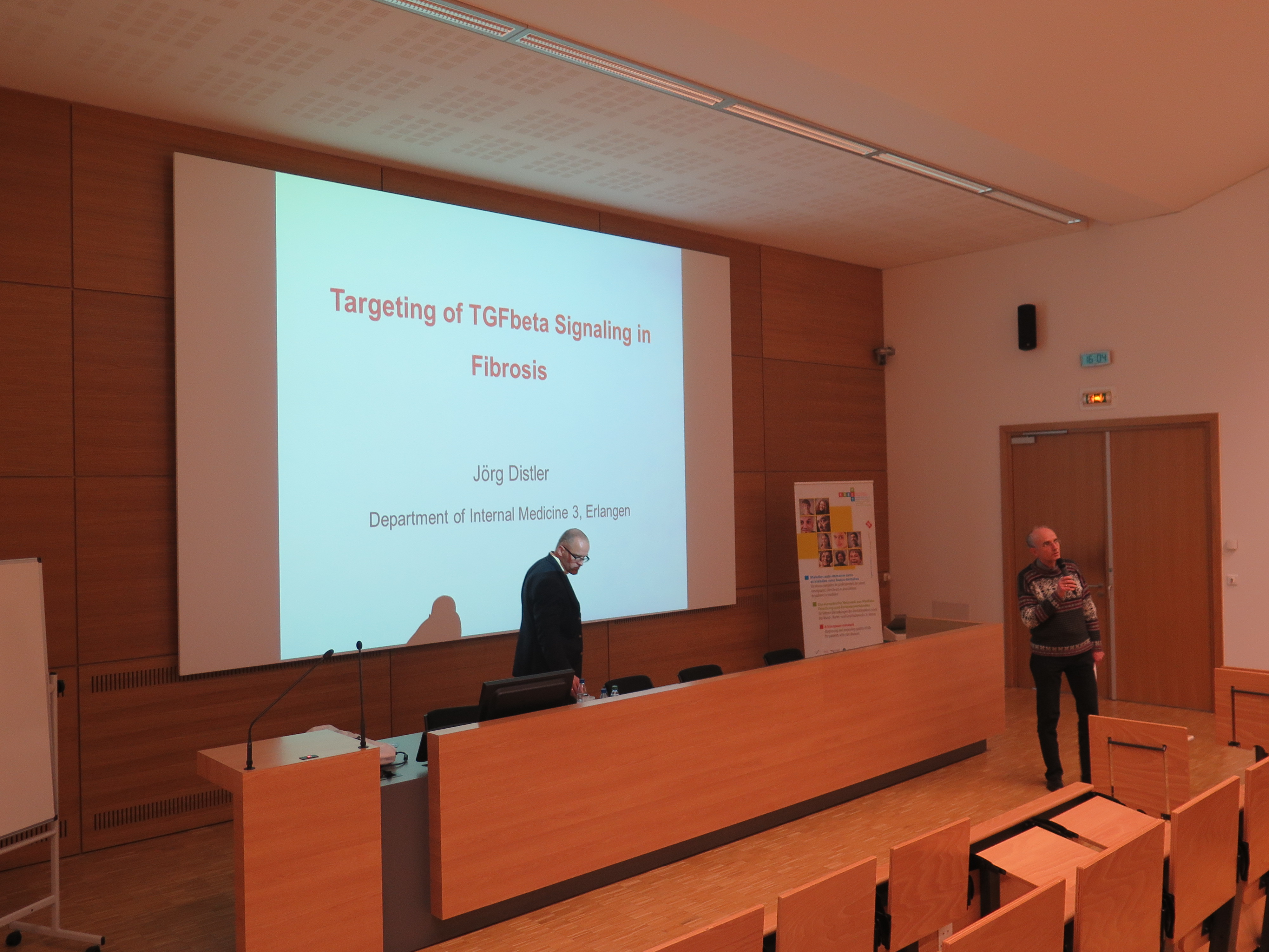
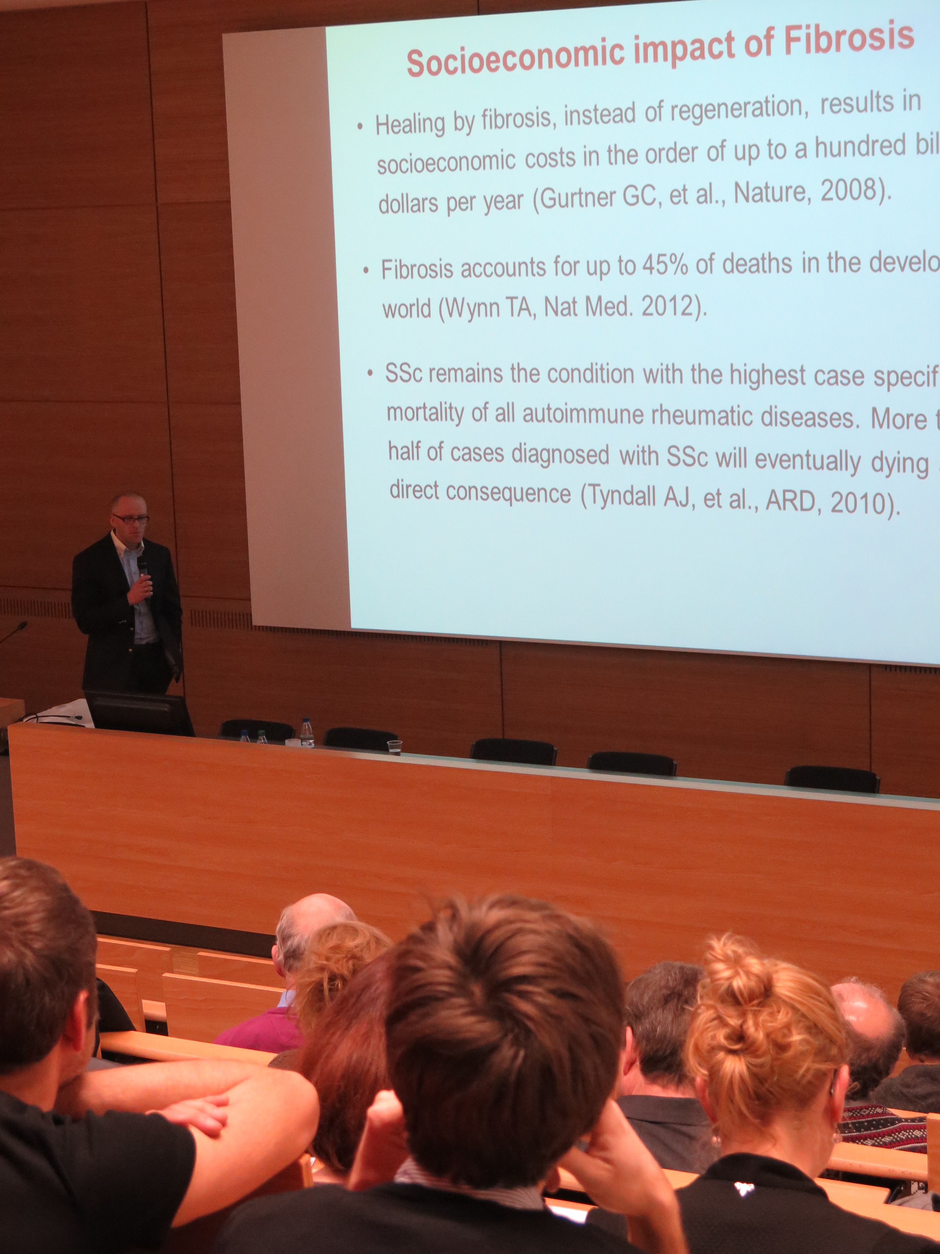
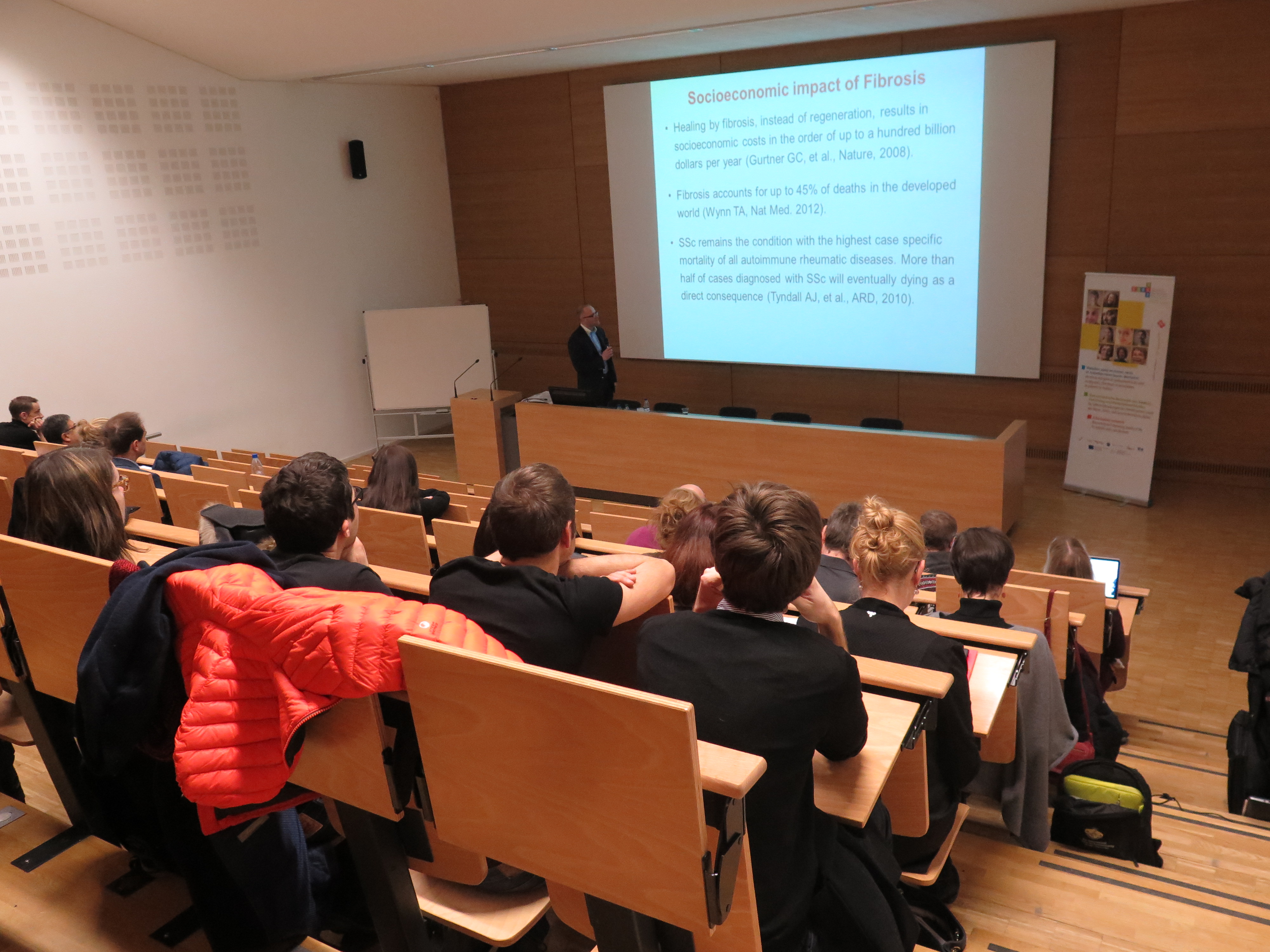
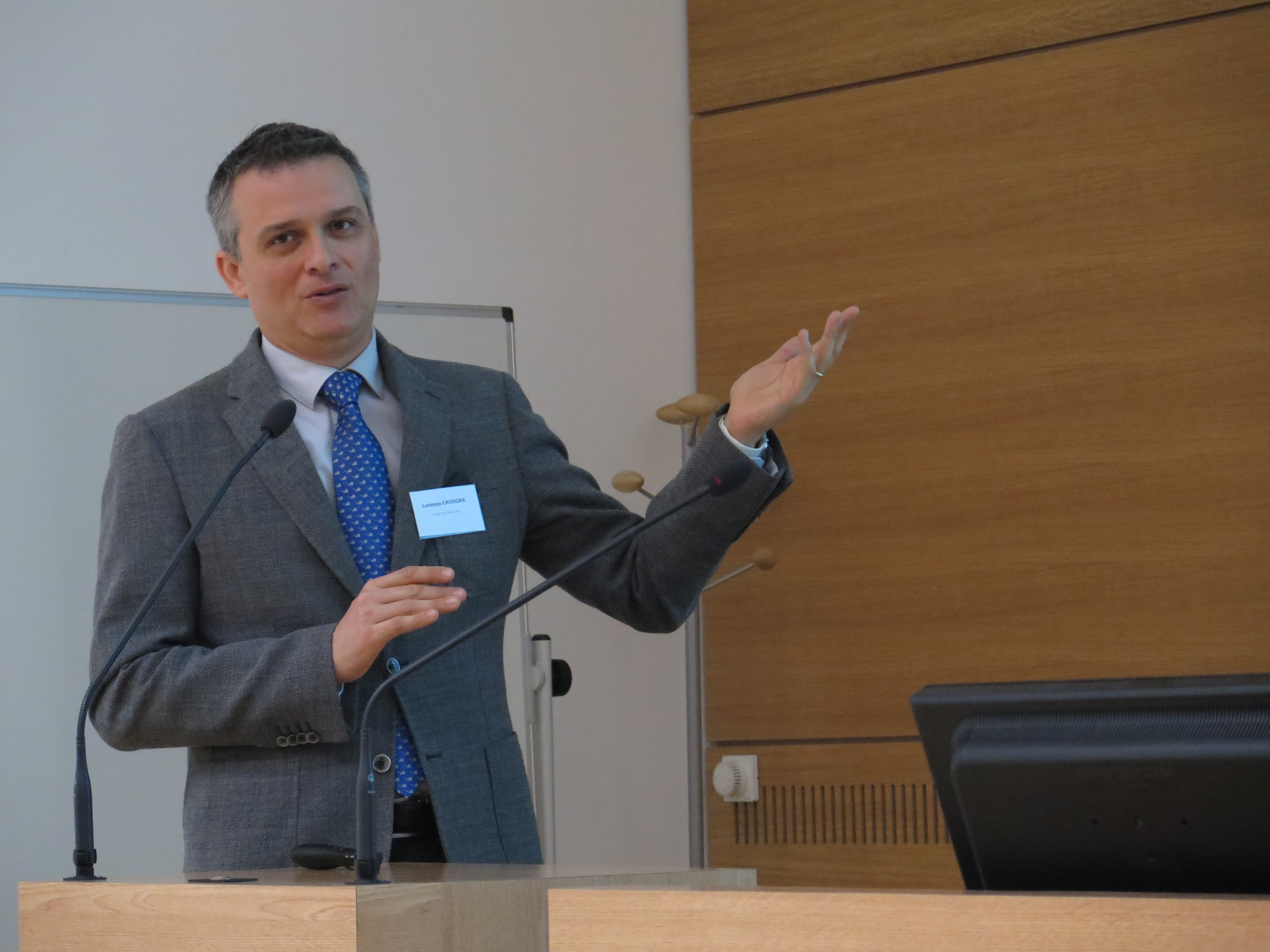
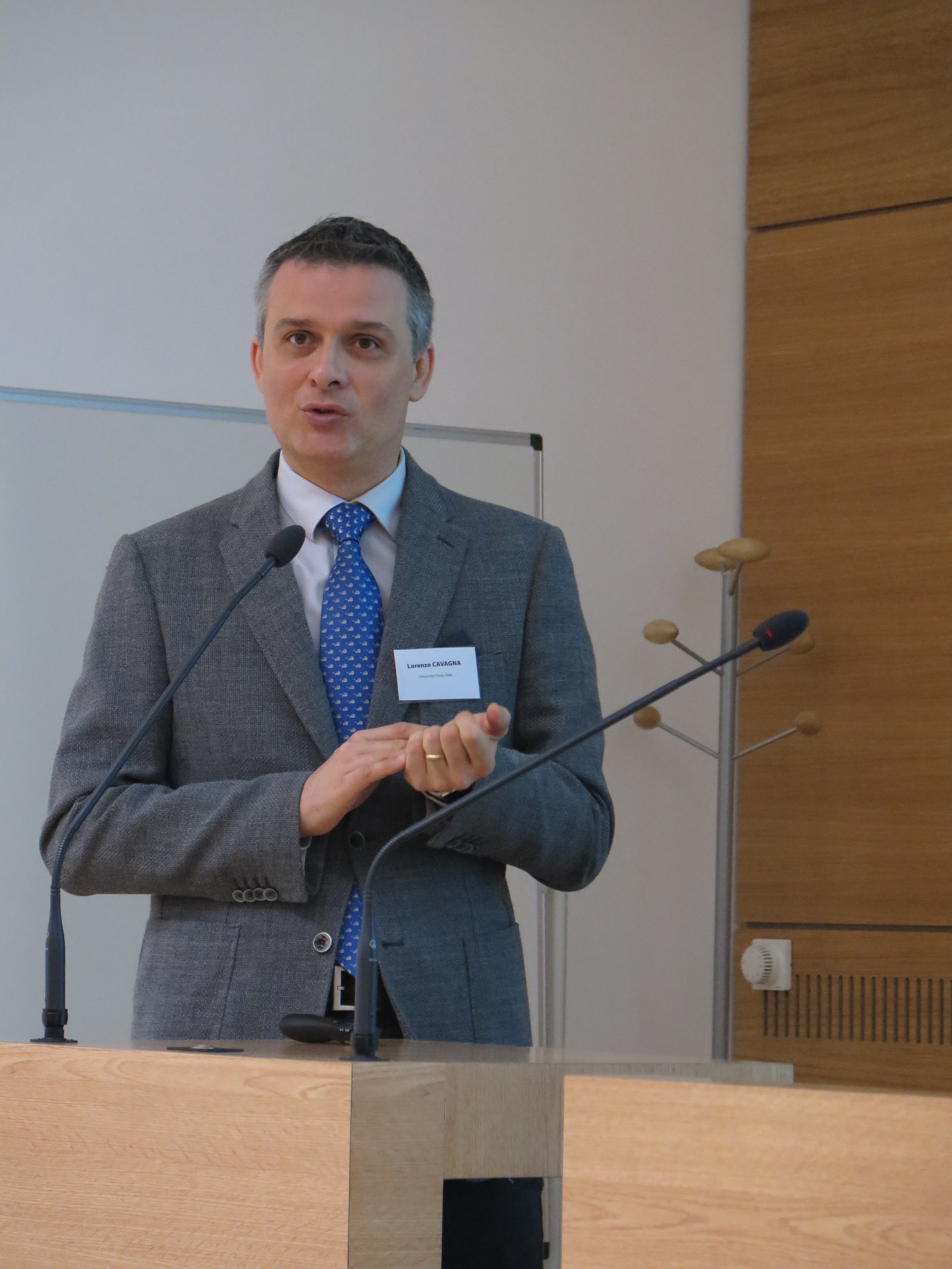
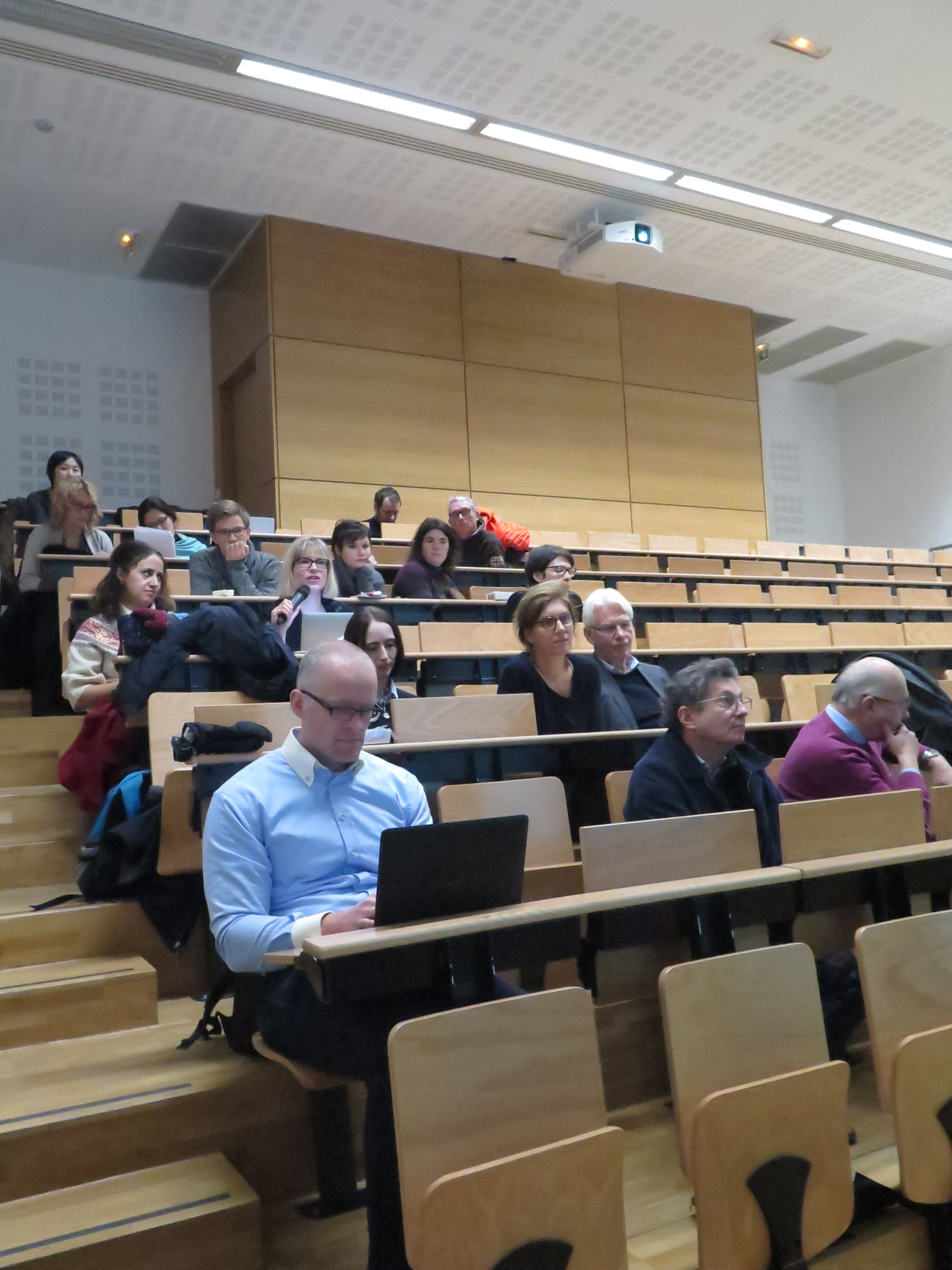
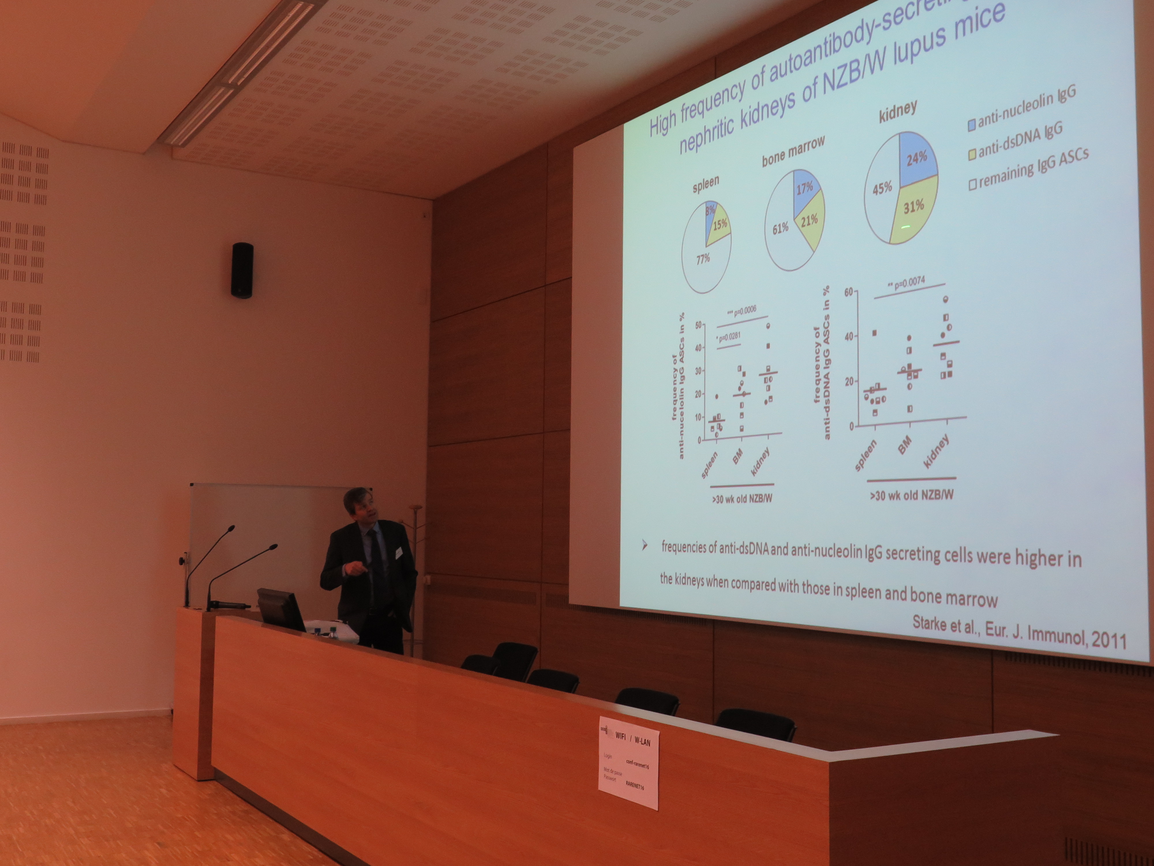
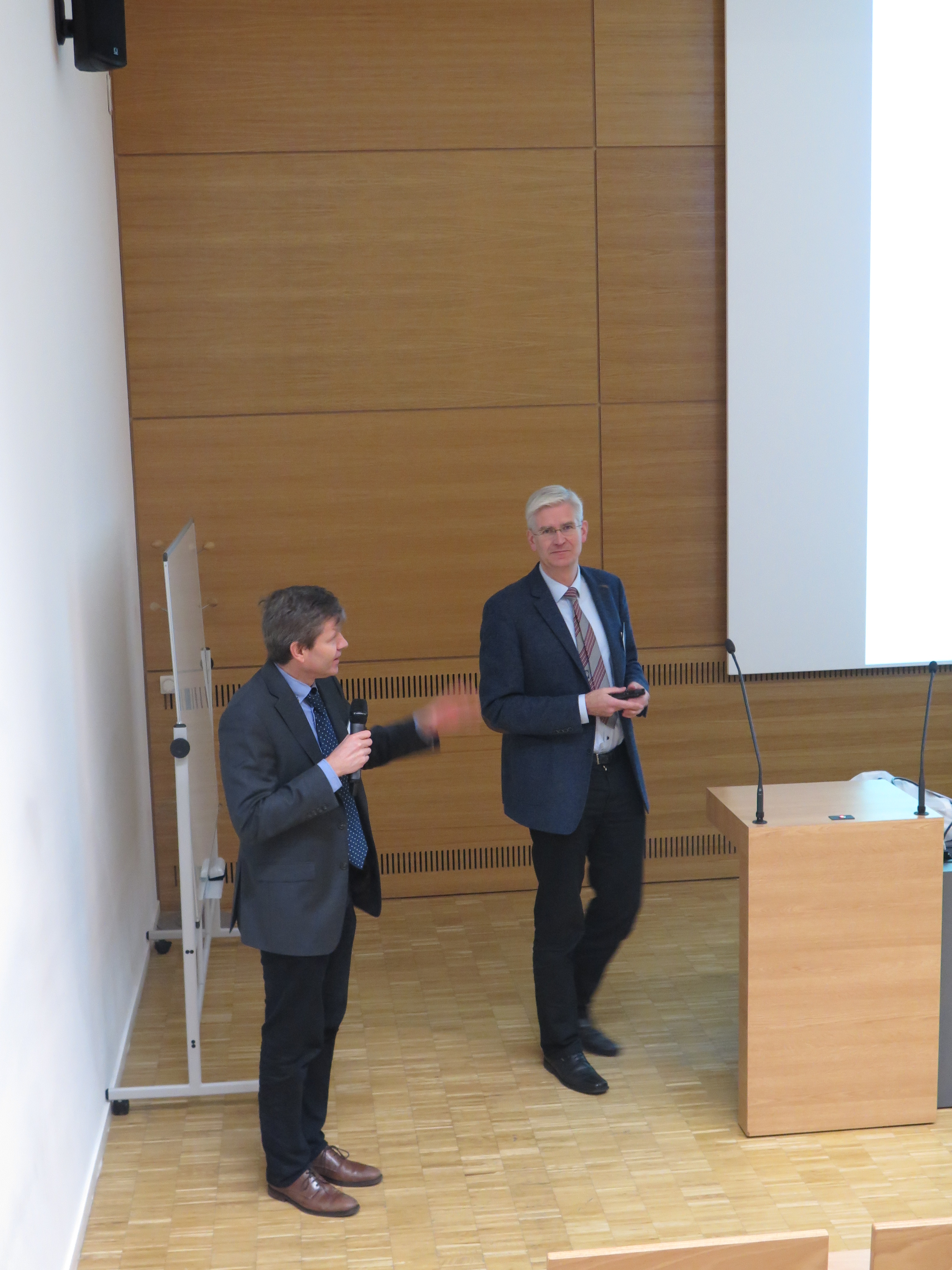
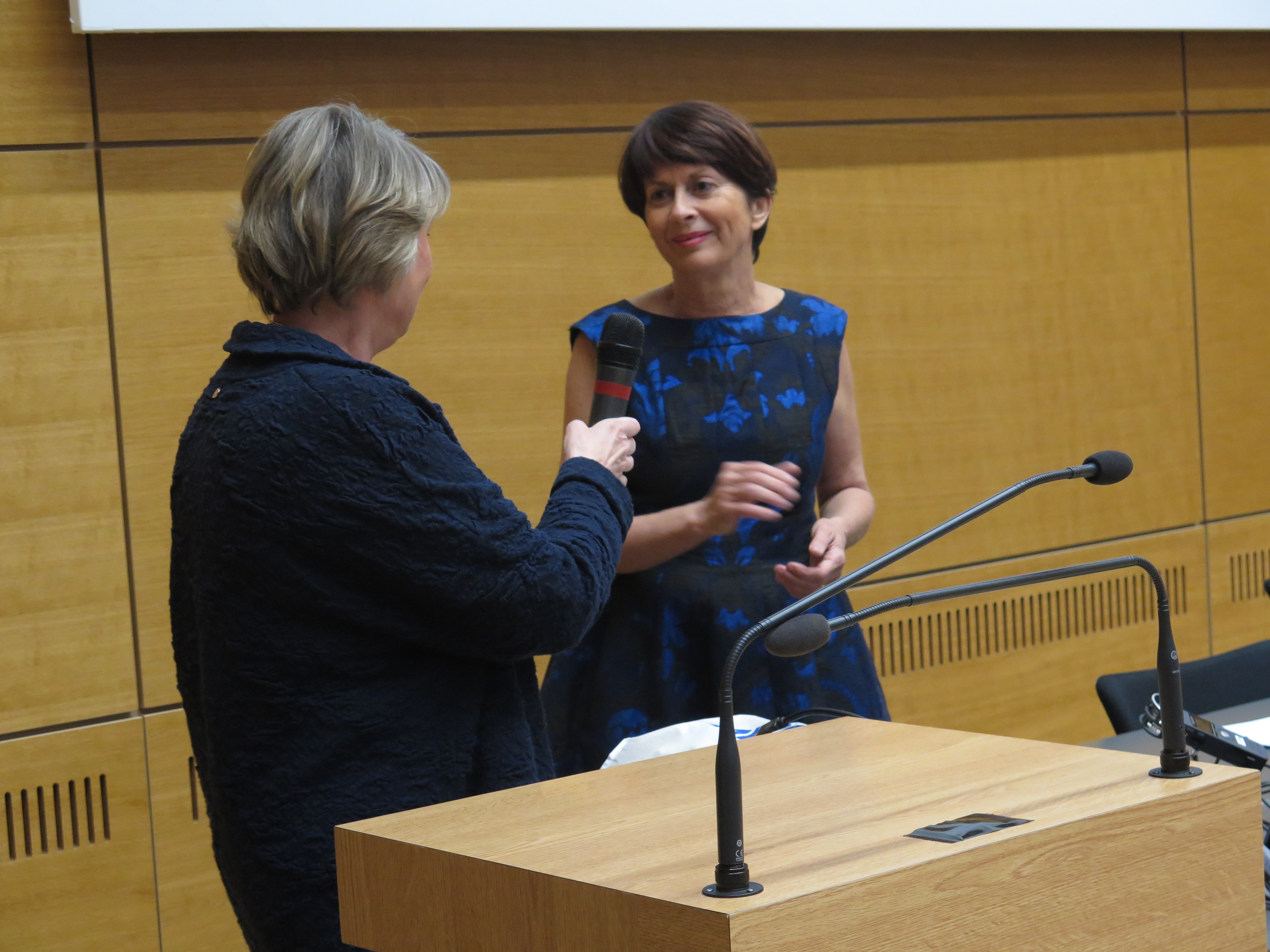
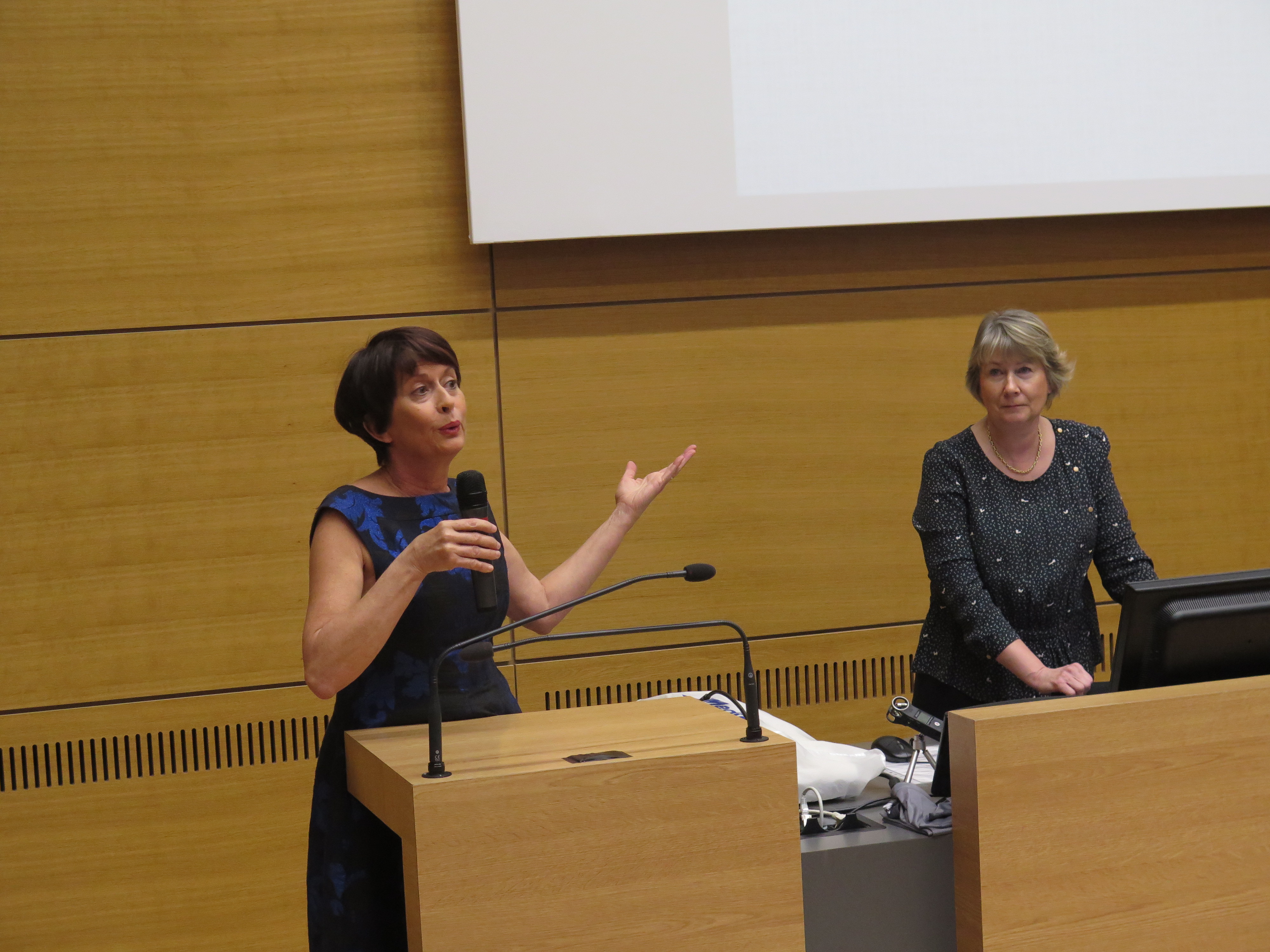
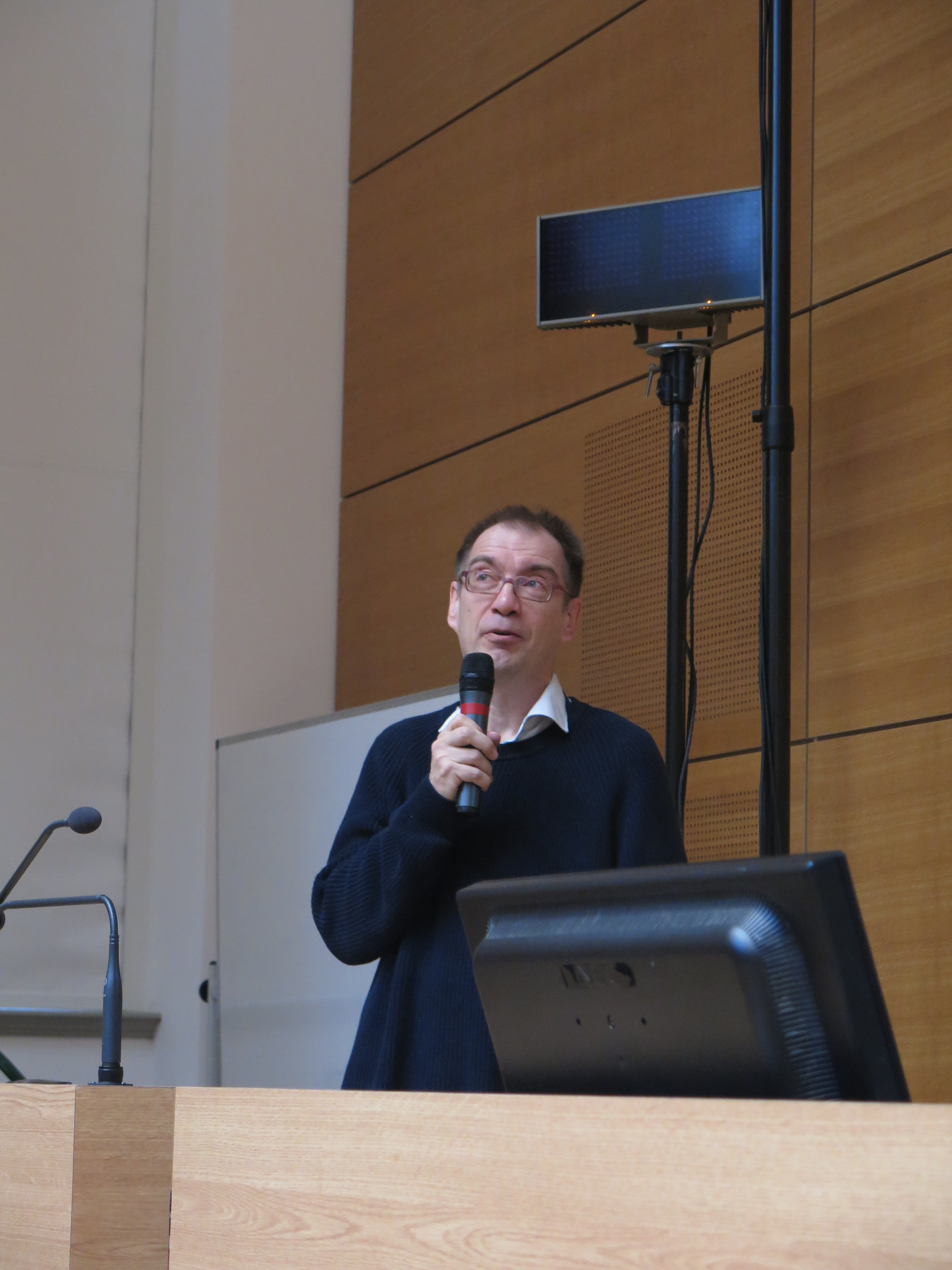
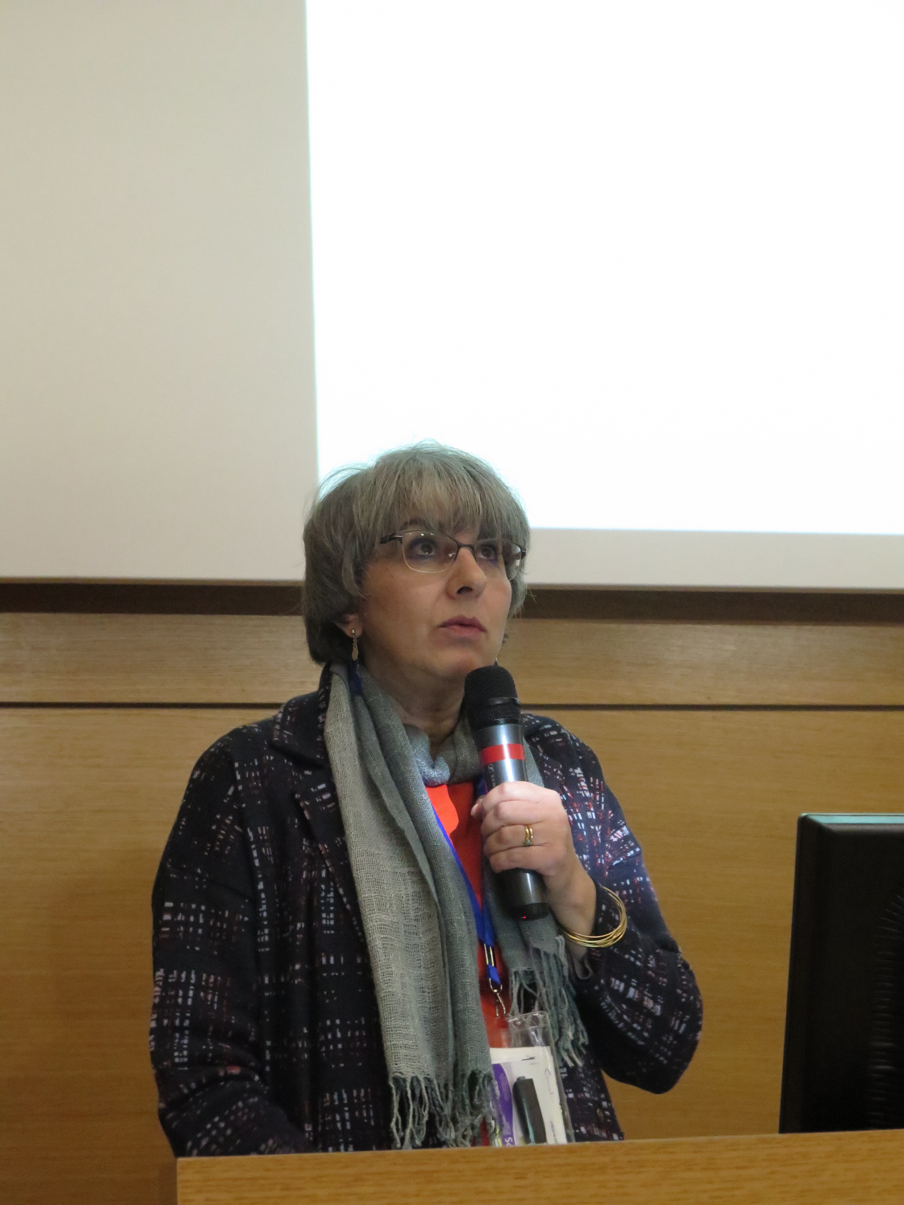
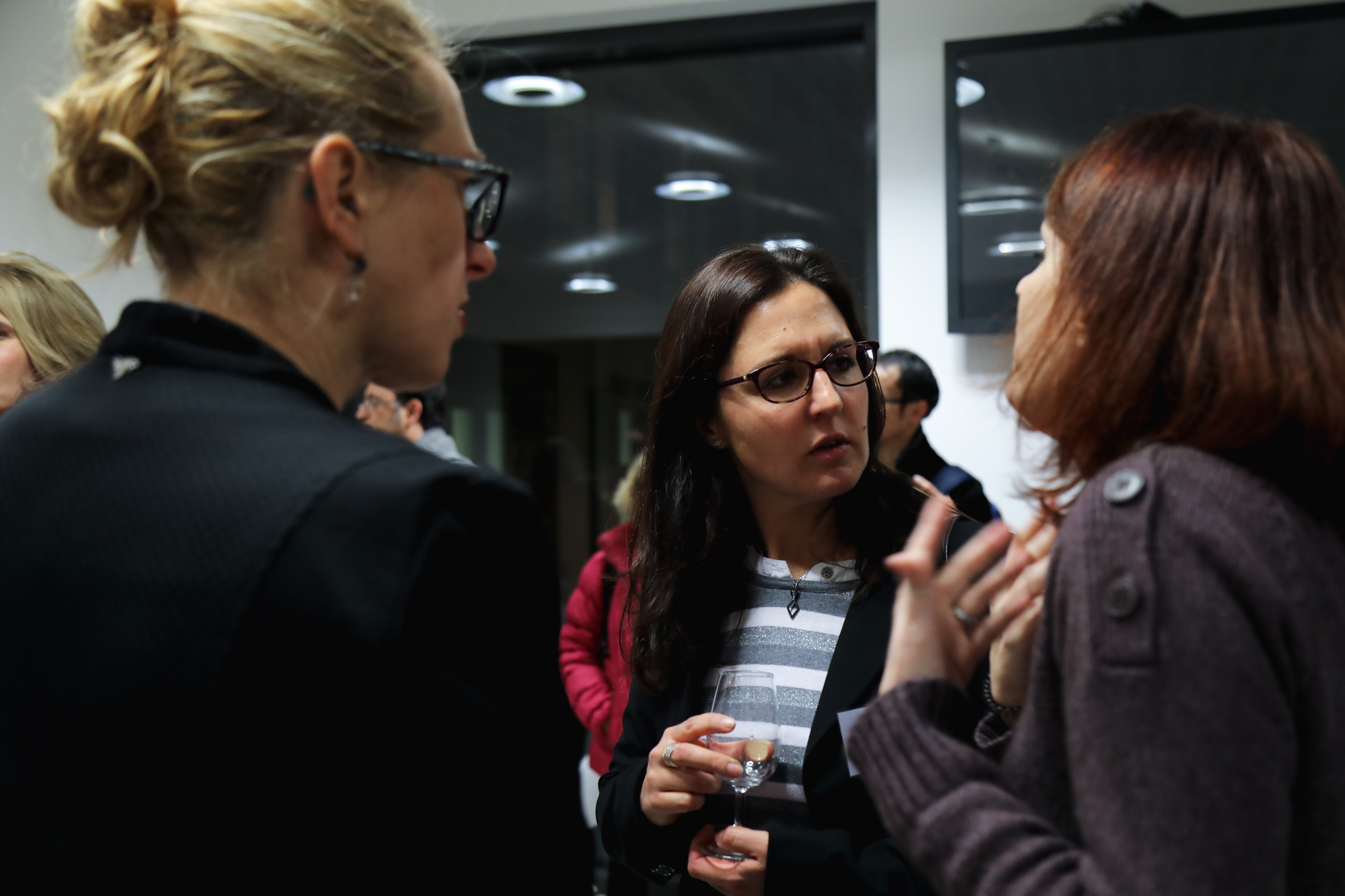
Le symposium “Maladies auto-immunes rares et vascularites” organisé dans le cadre du projet RARENET a réuni une quarantaine de participants les mercredi 7 et jeudi 8 décembre à l’Université de Strasbourg. Grâce à la présence d’experts français, allemands et italiens, le programme scientifique était de très grande qualité.
L’après-midi du 8 décembre a été consacrée à un échange avec les associations de patients : Associations des Sclérodermiques de France (ASF) et Association française du lupus et autre maladies auto-immmunes (AFL).
Retour en images:
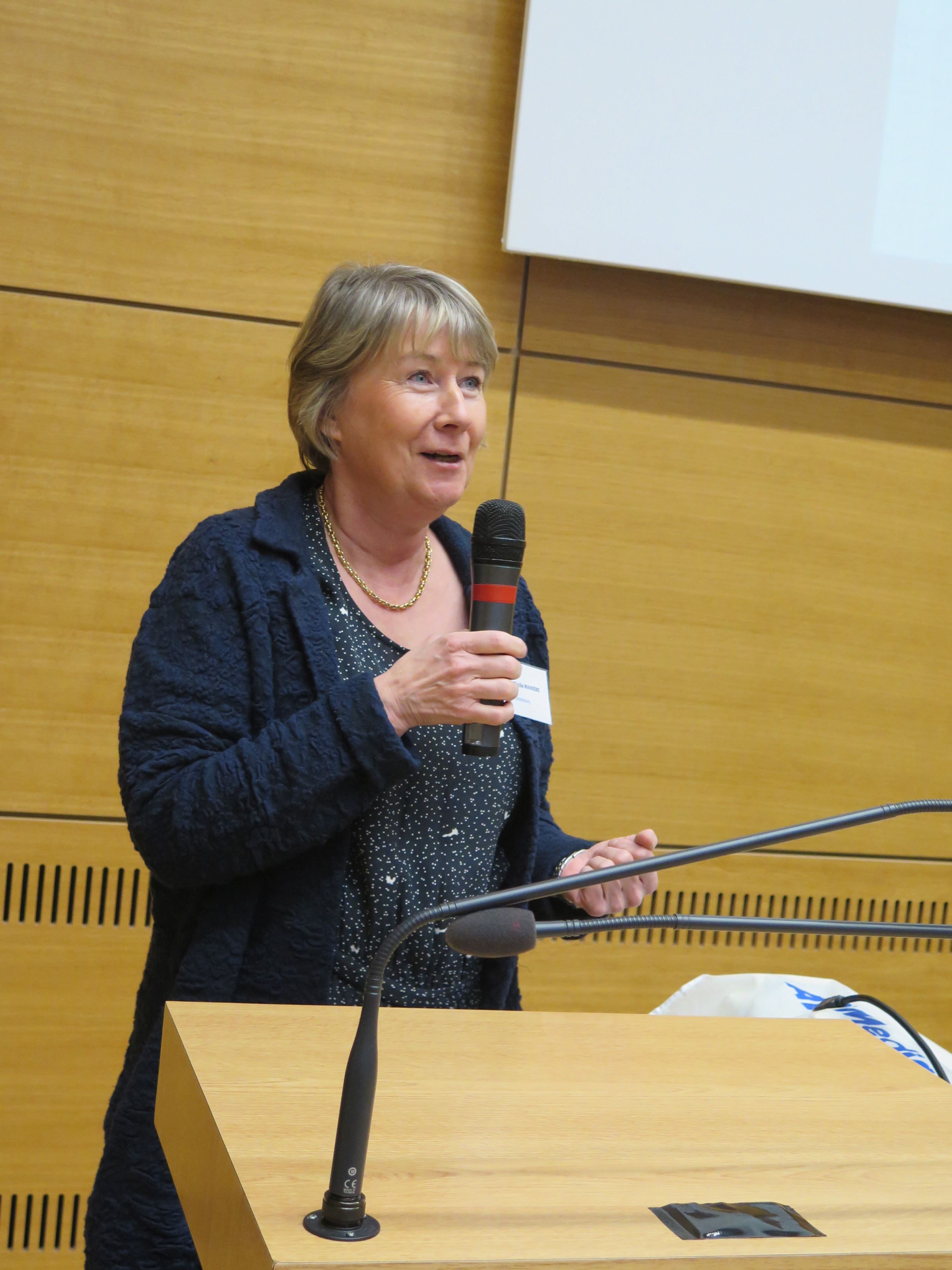
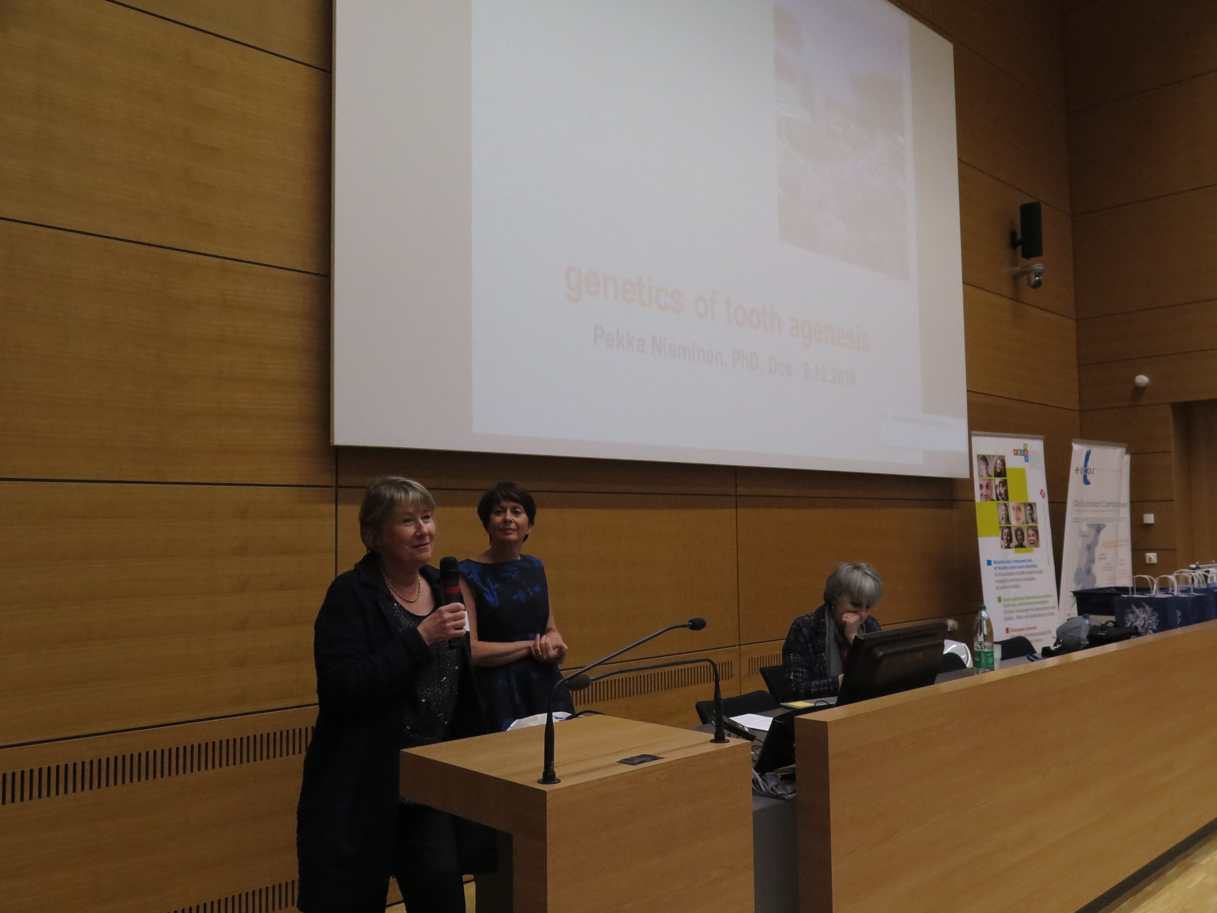
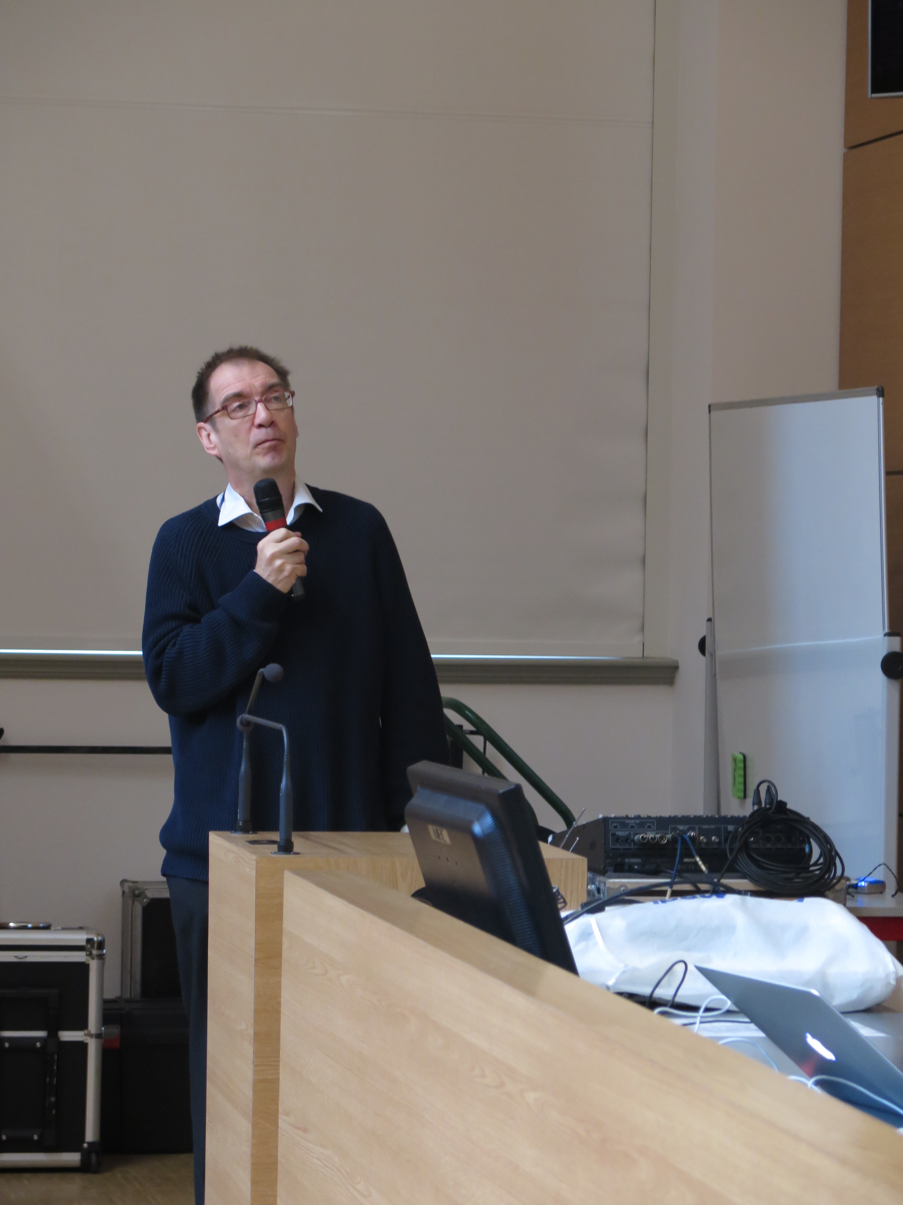
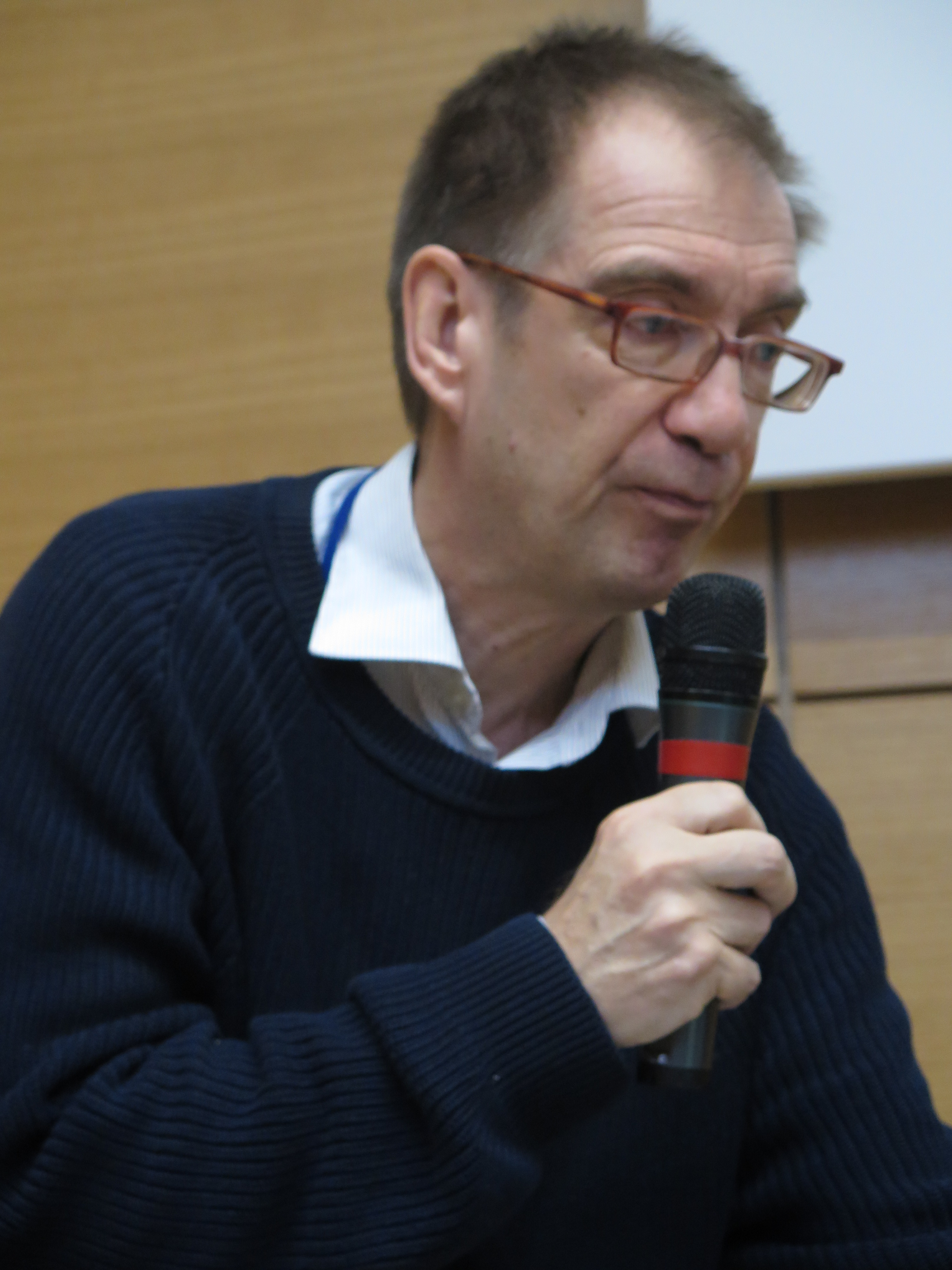
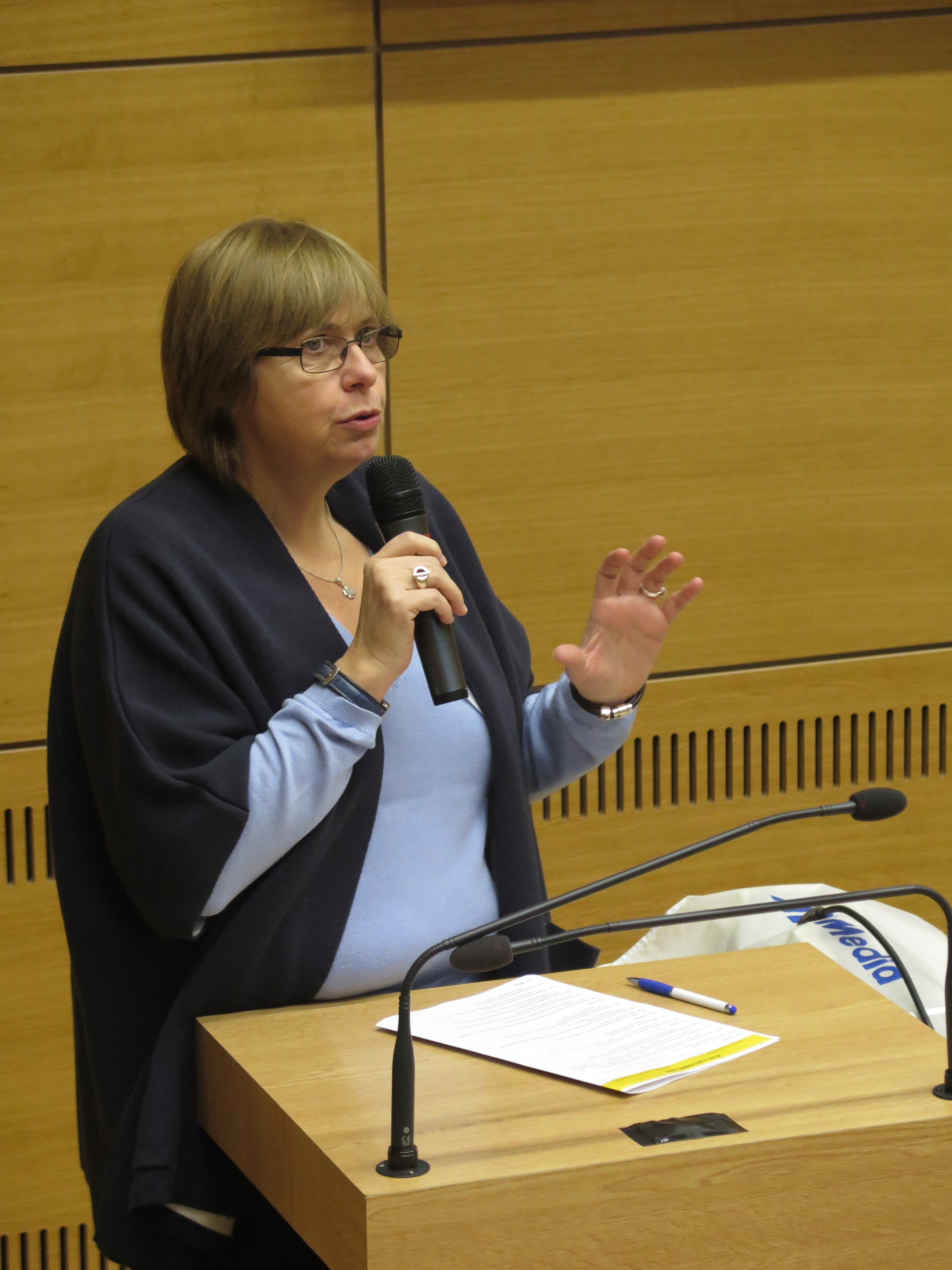
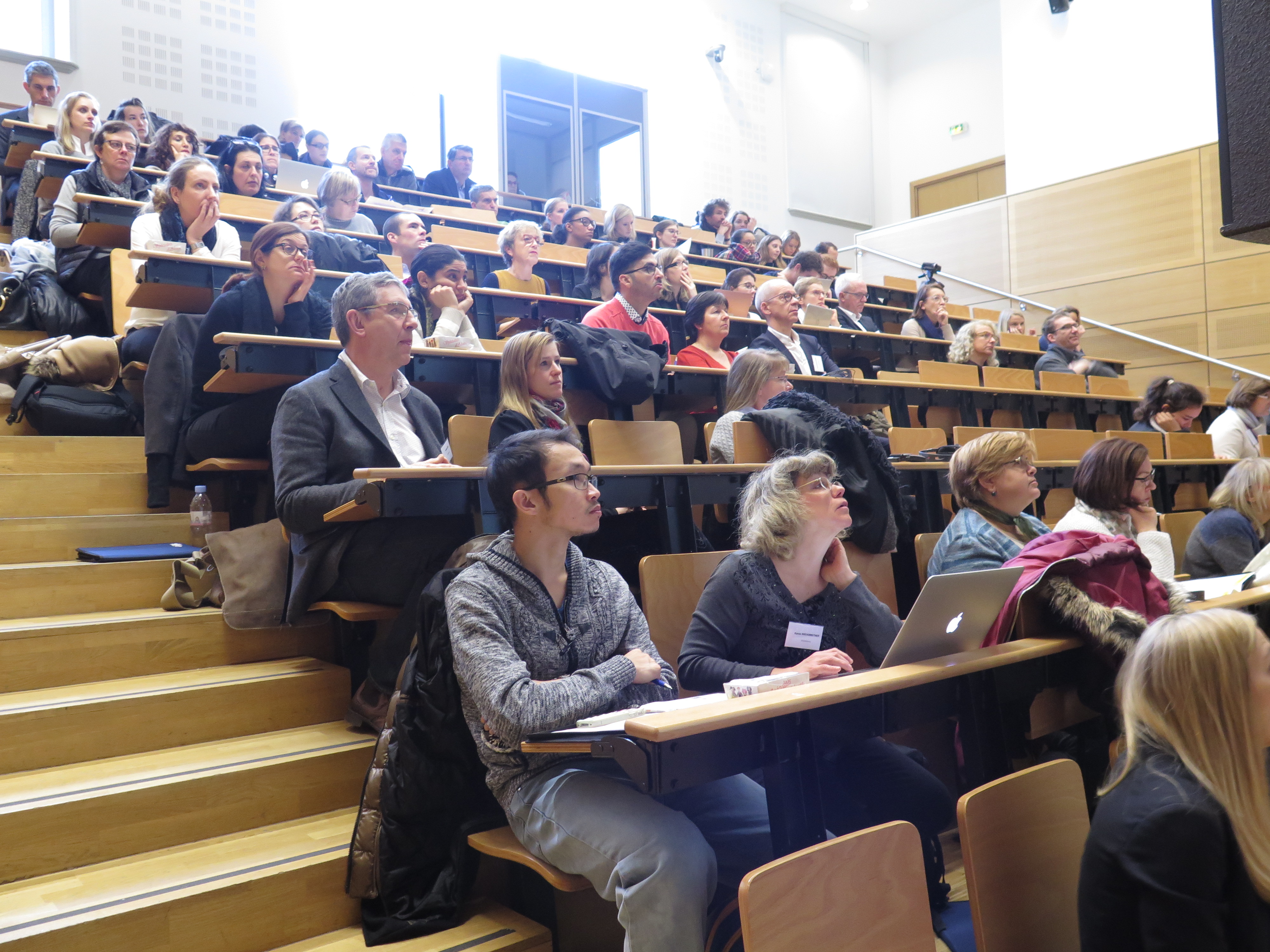
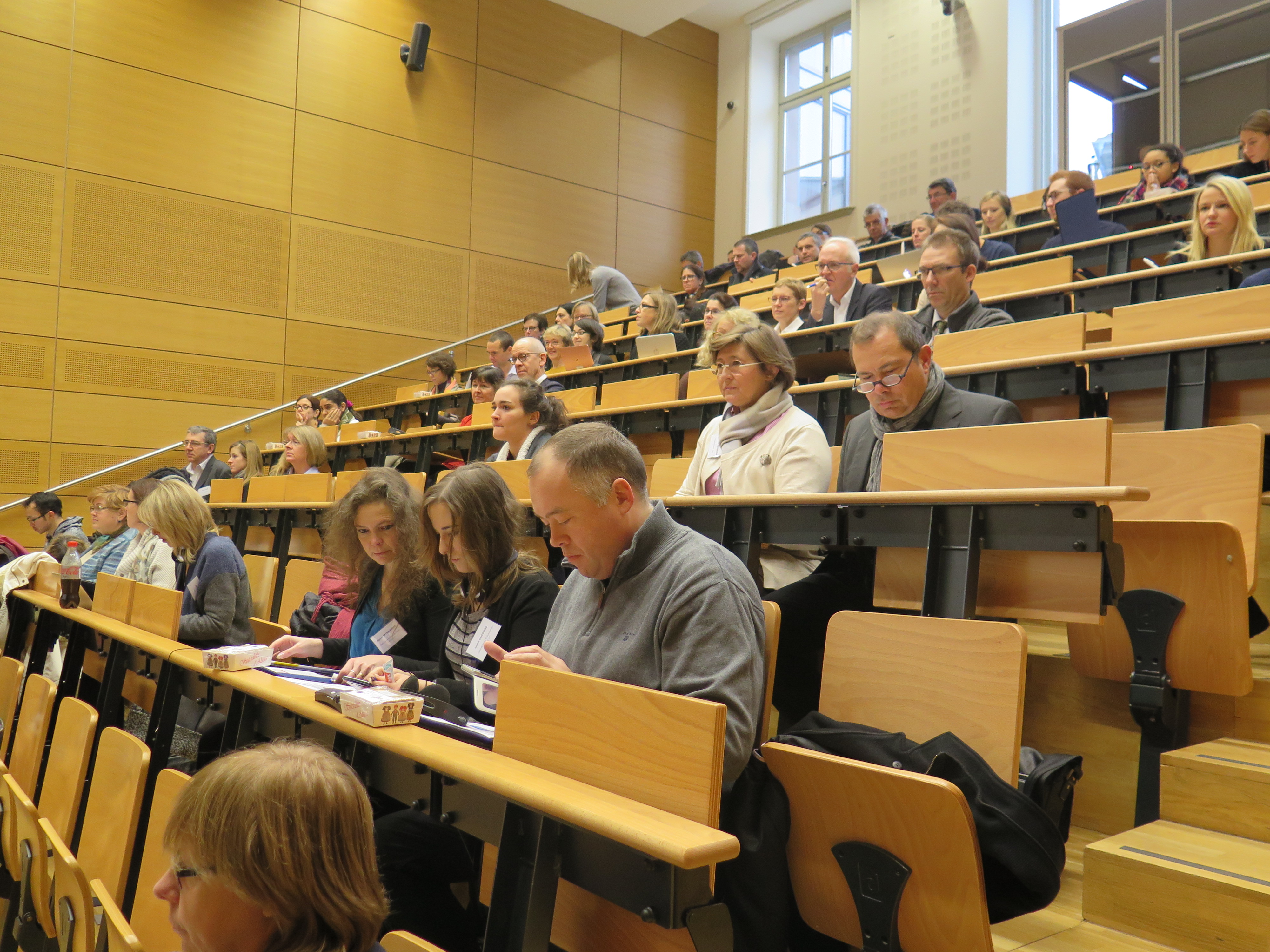
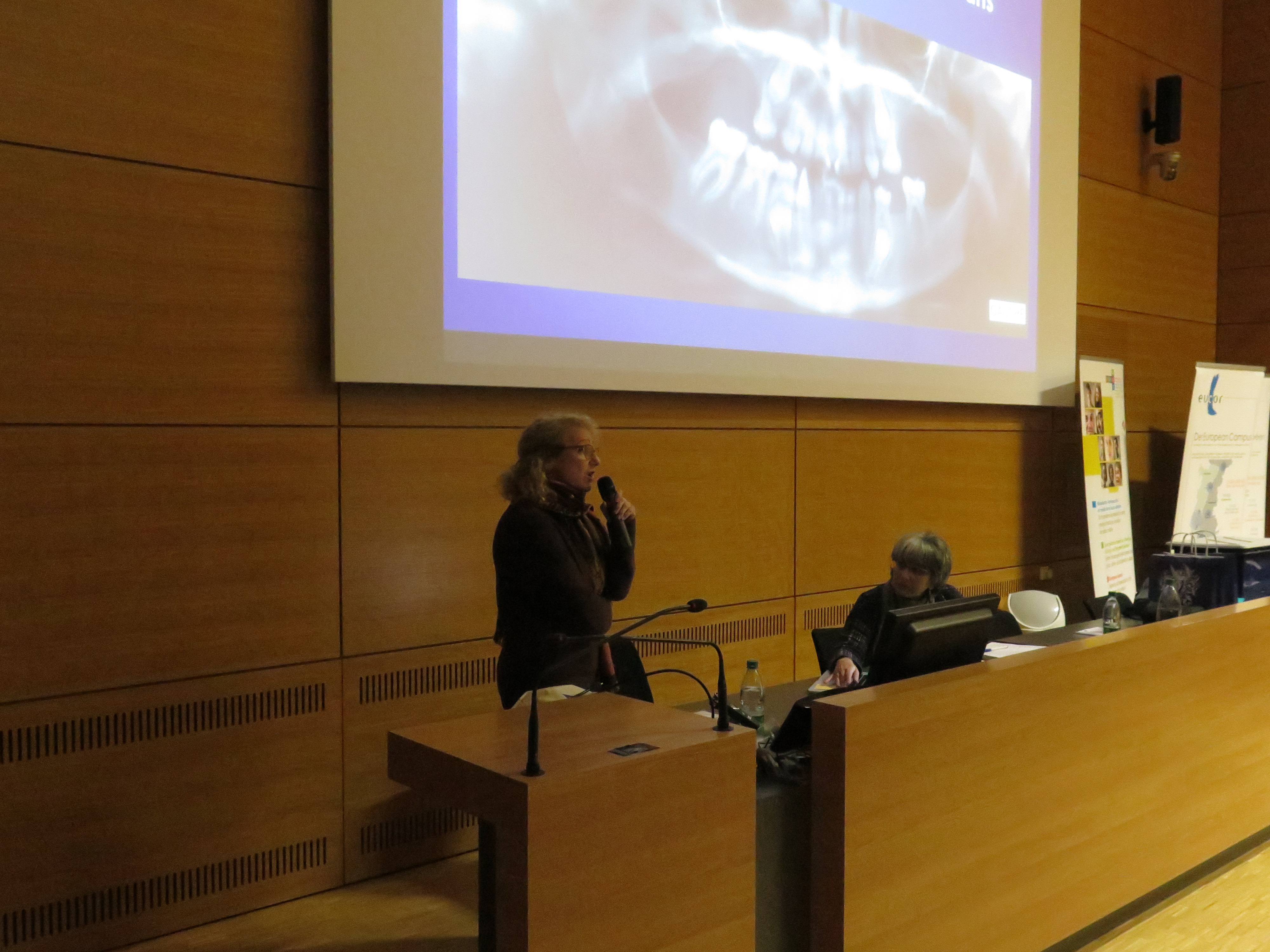
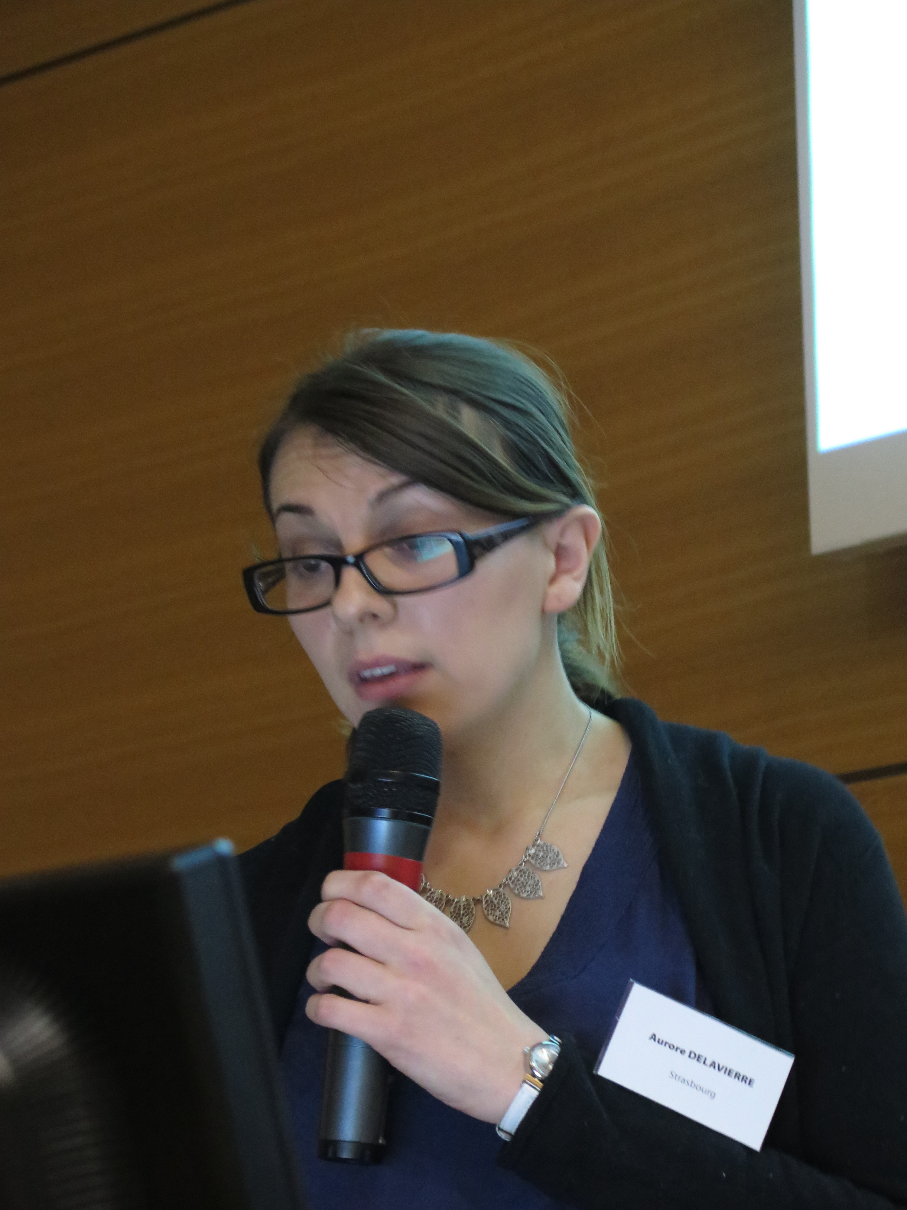
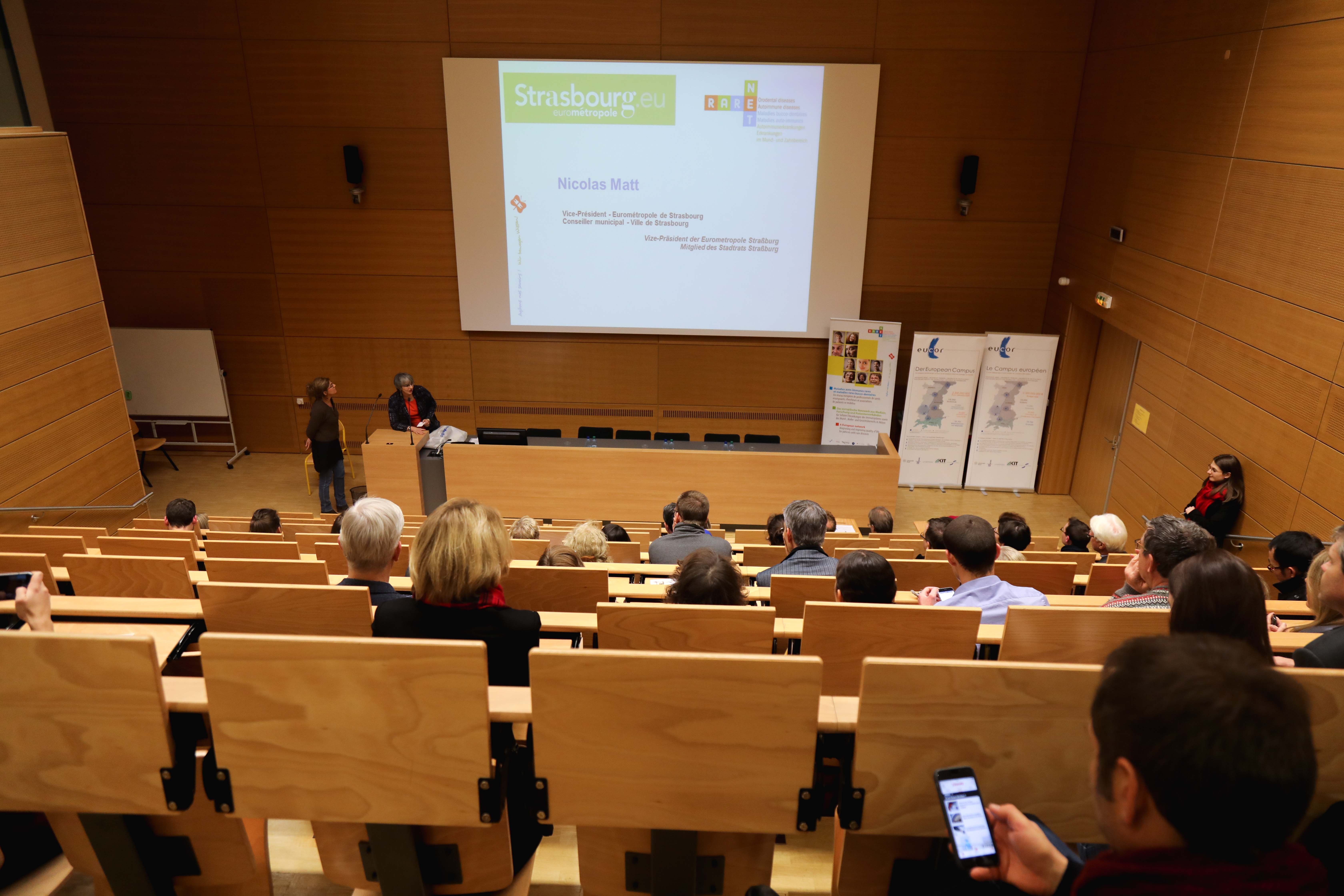
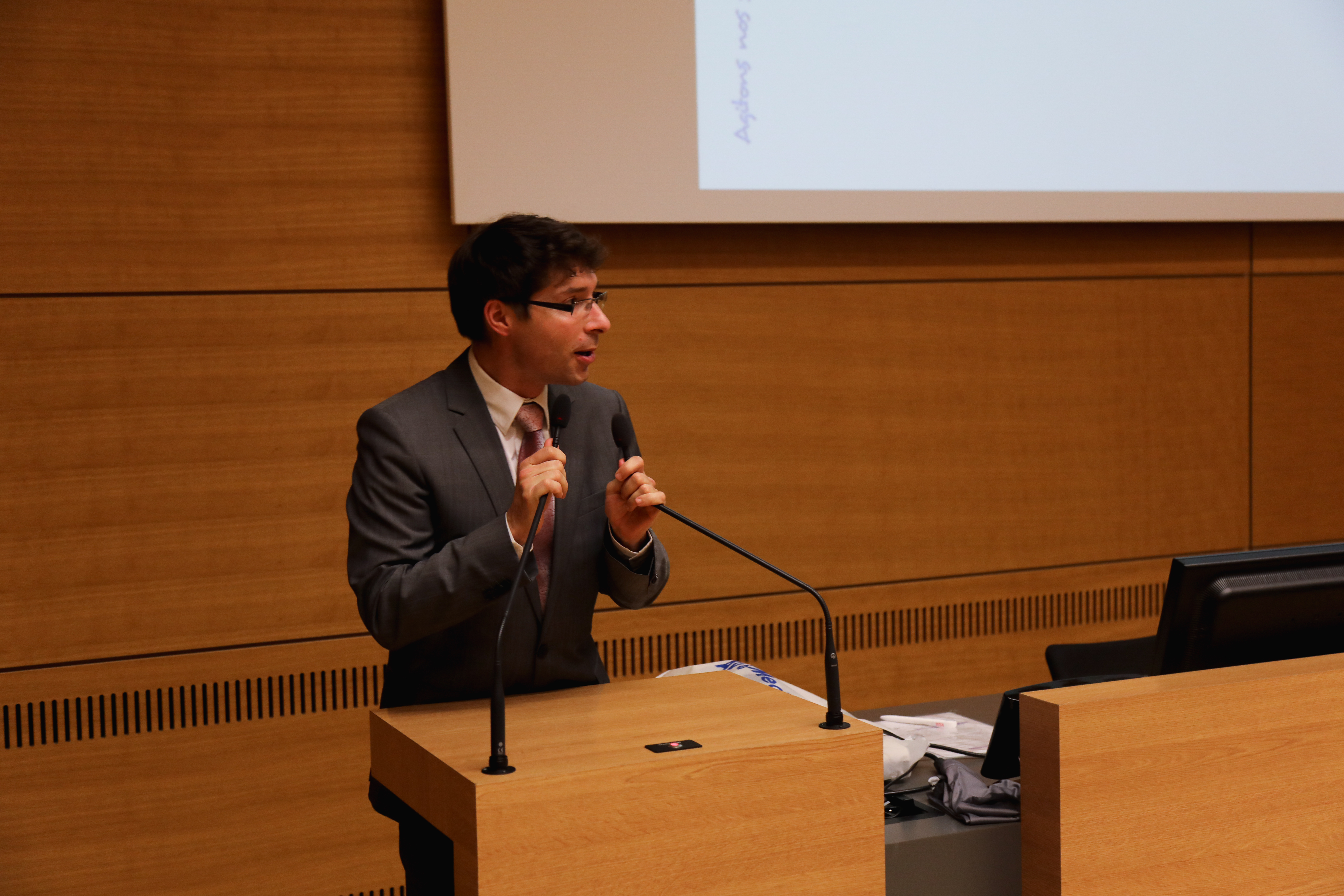
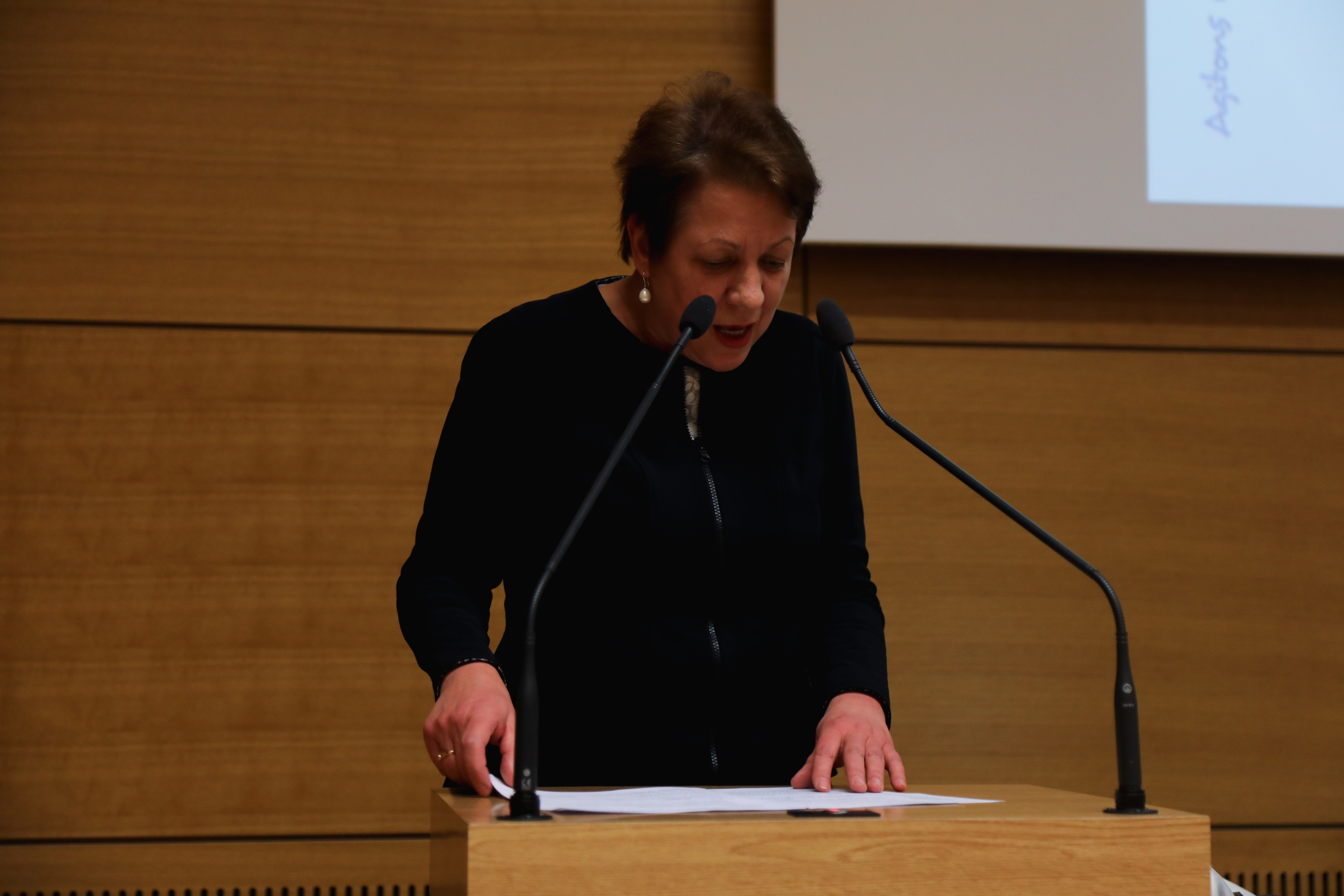
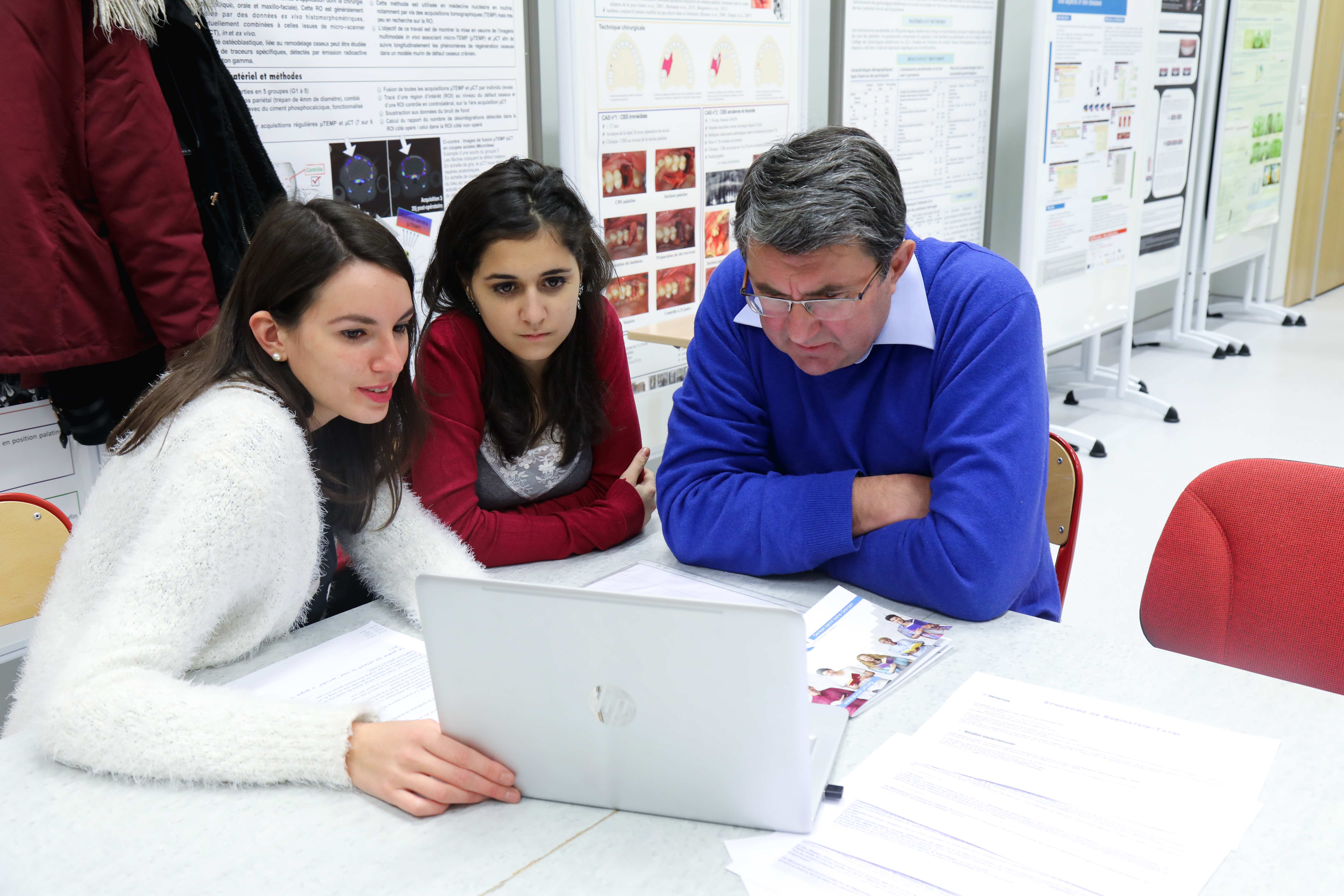
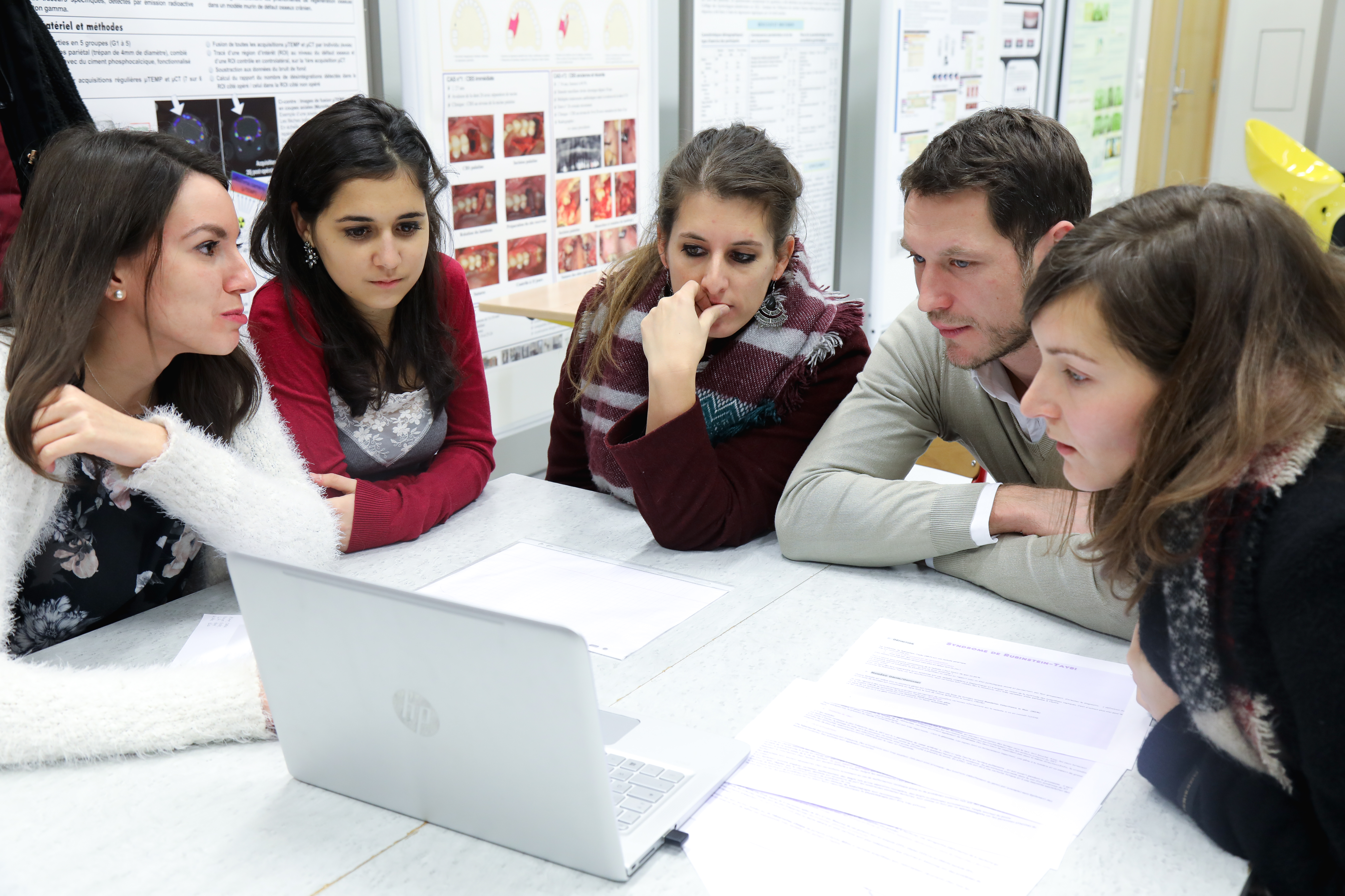
Le symposium “Agénésies dentaires multiples”, organisé à l’occasion du lancement de RARENET et des 10 ans du Centre de Référence O-Rares, a réuni près de 80 participants le jeudi 8 décembre à la Faculté de Chirurgie Dentaire de l’Université de Strasbourg.
Retour en images :
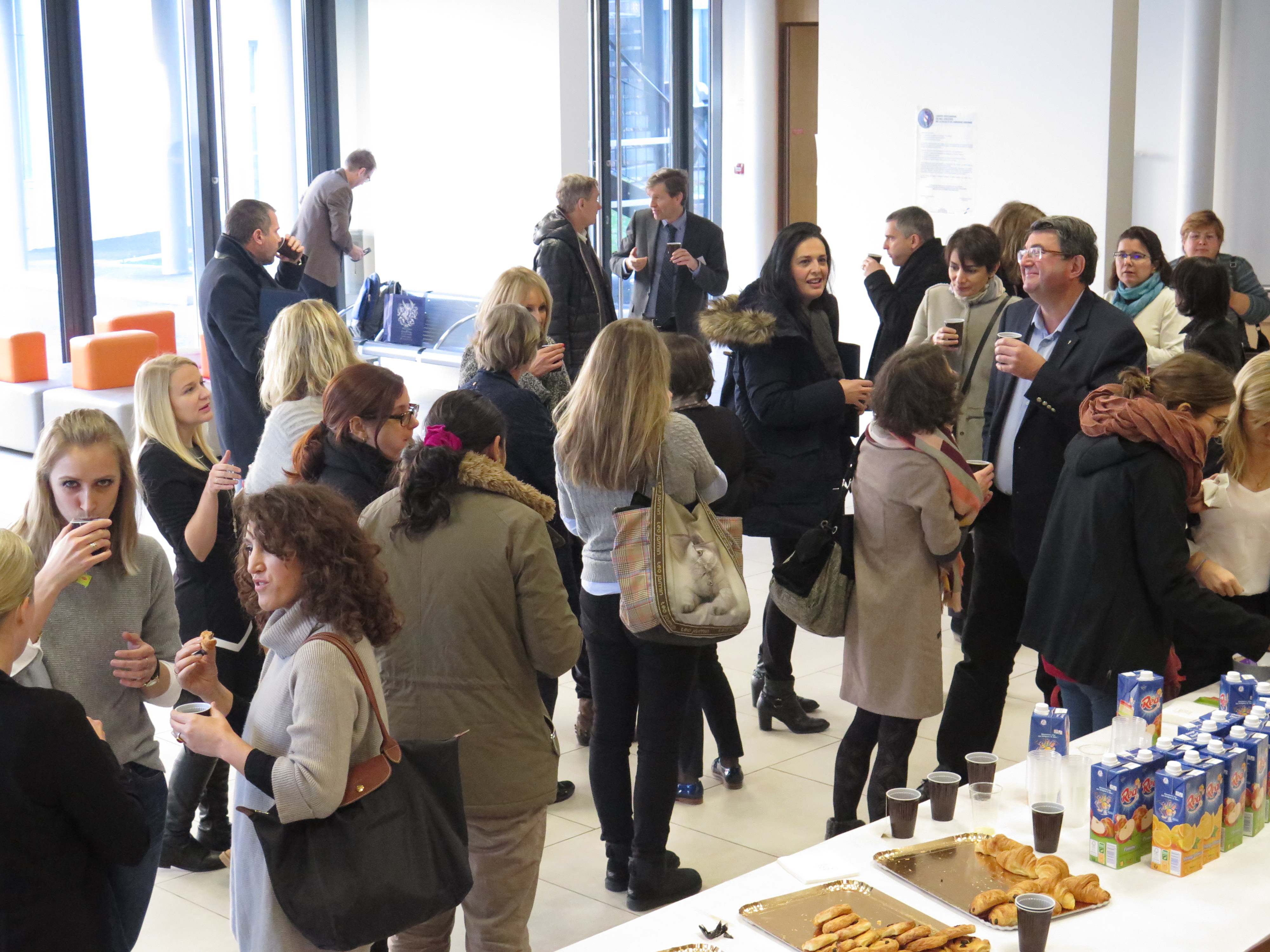
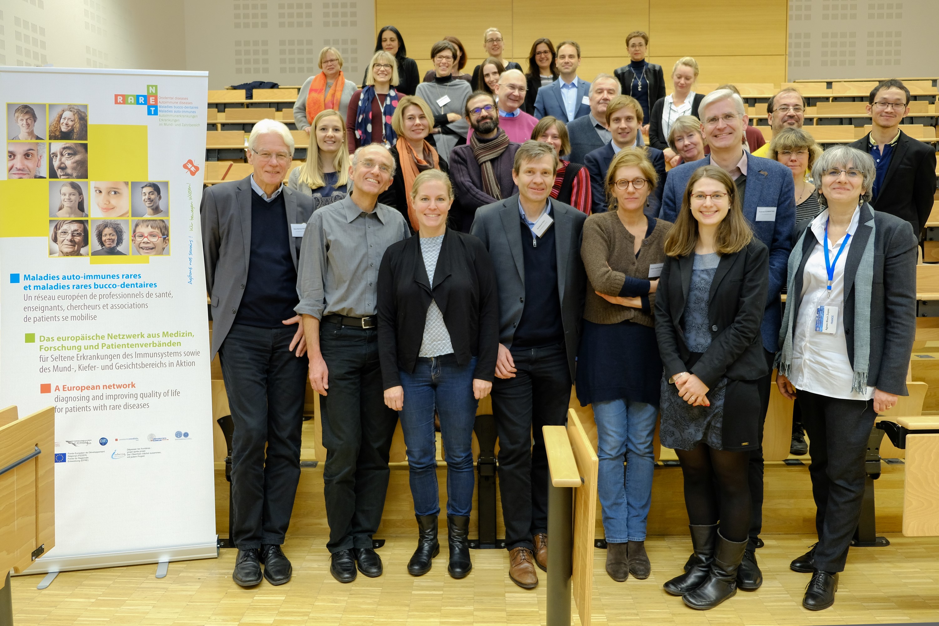
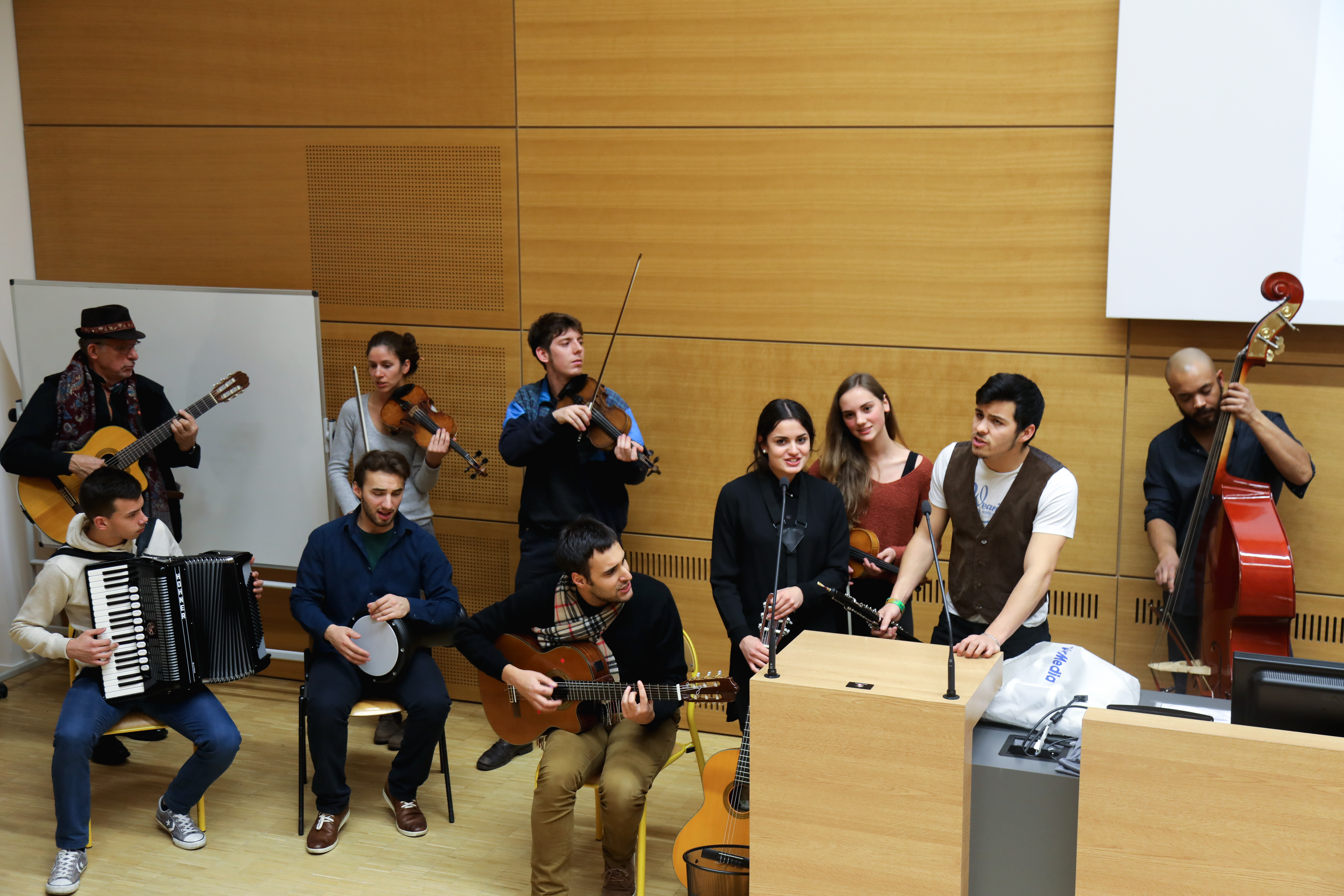
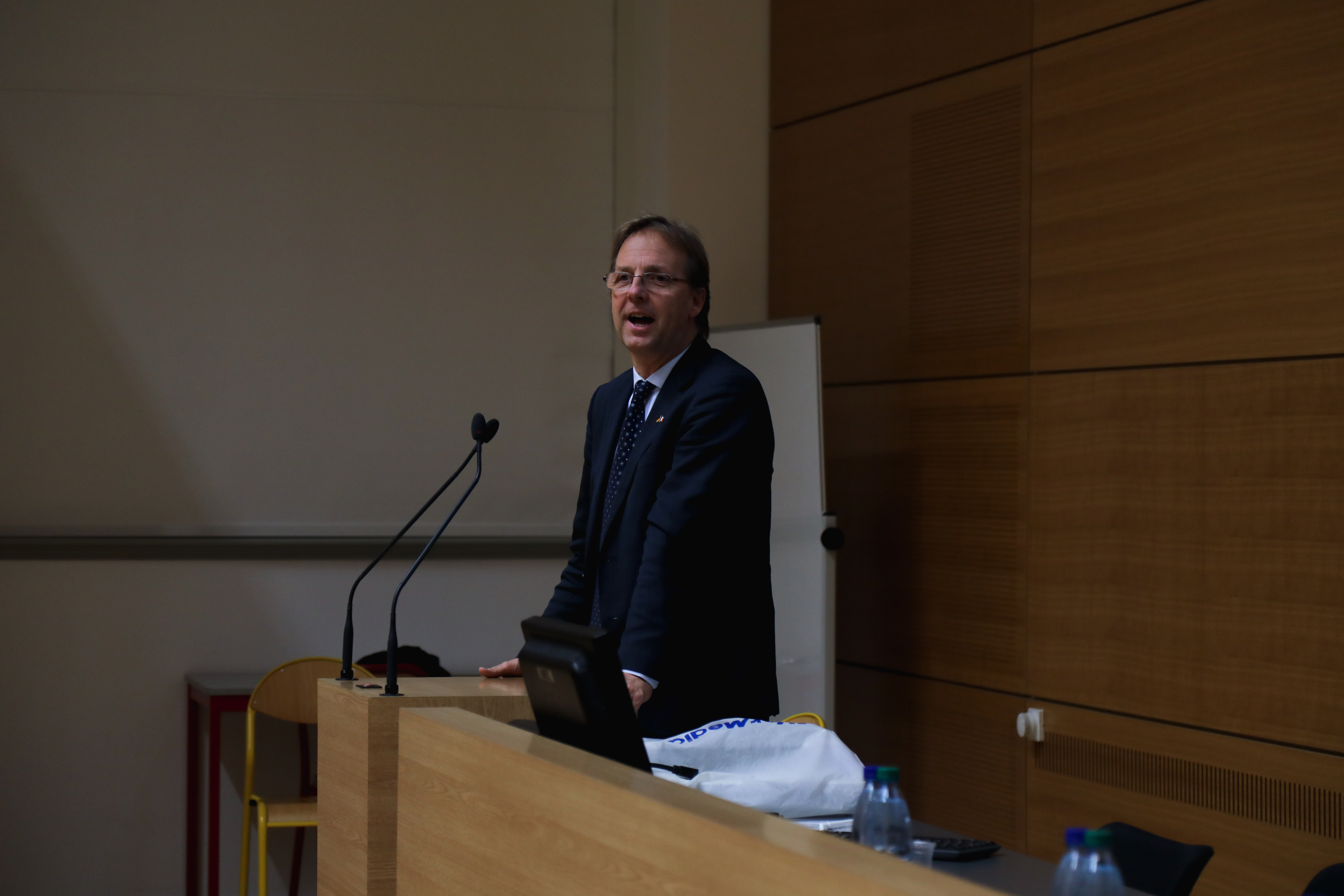
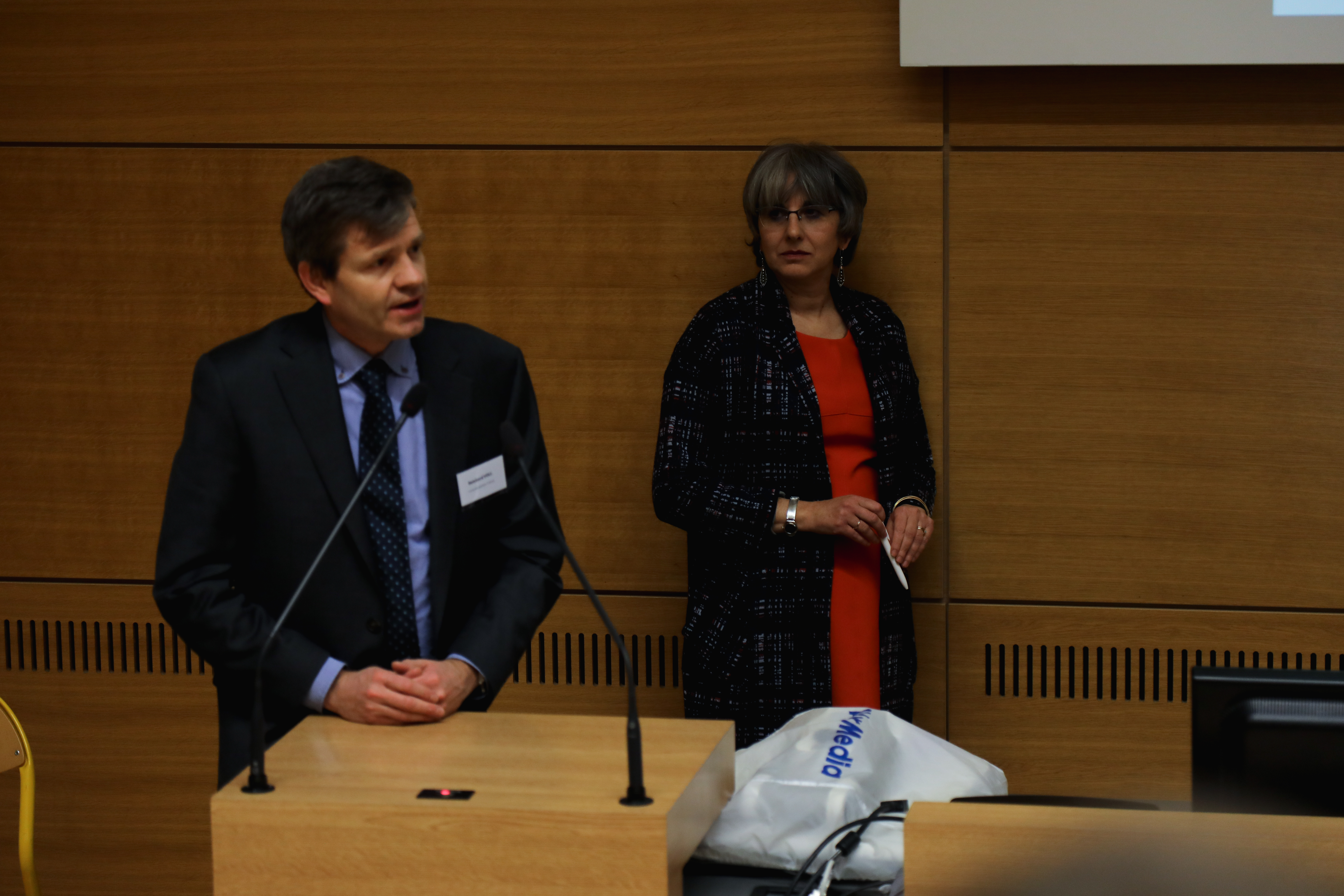
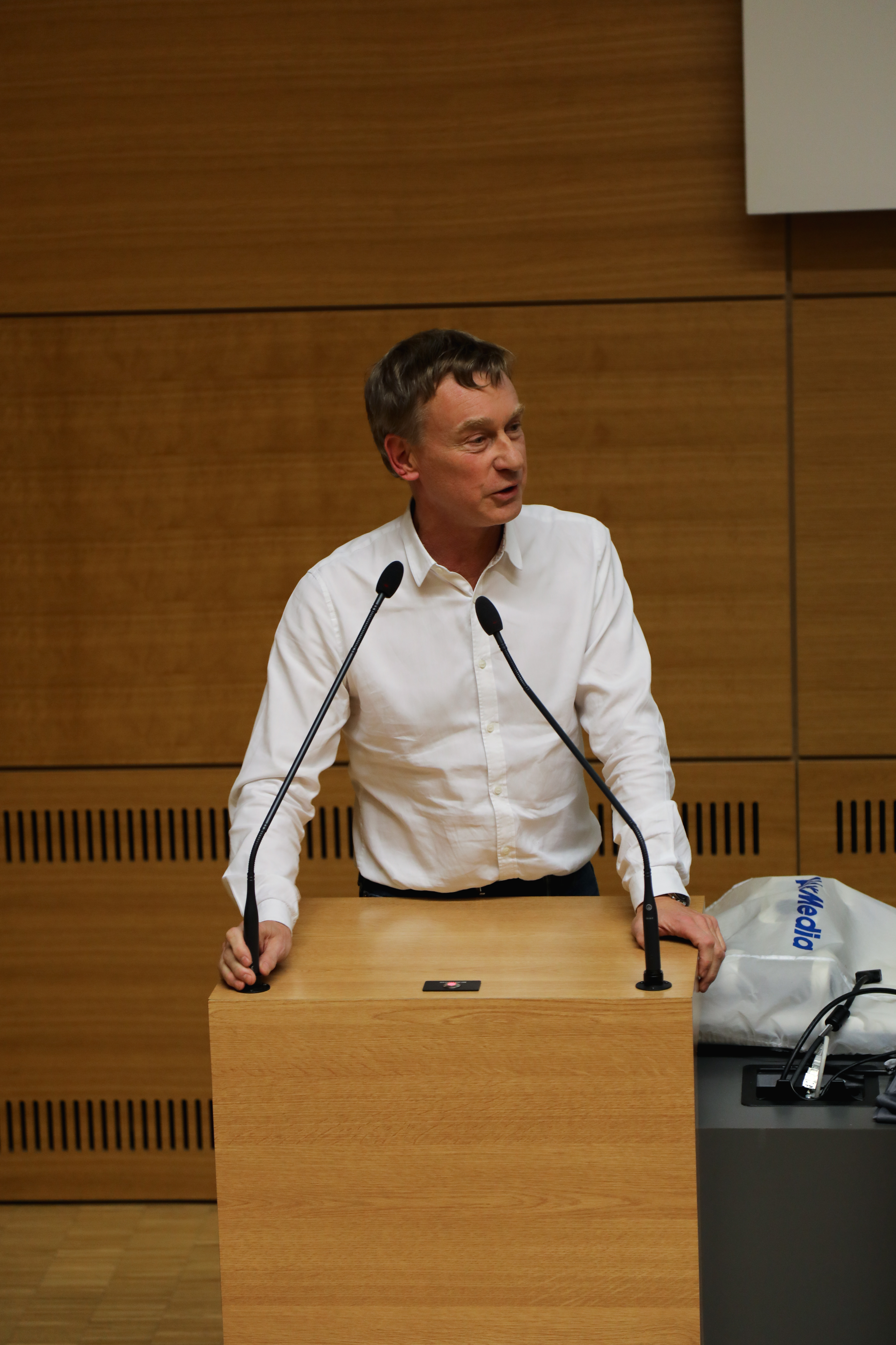
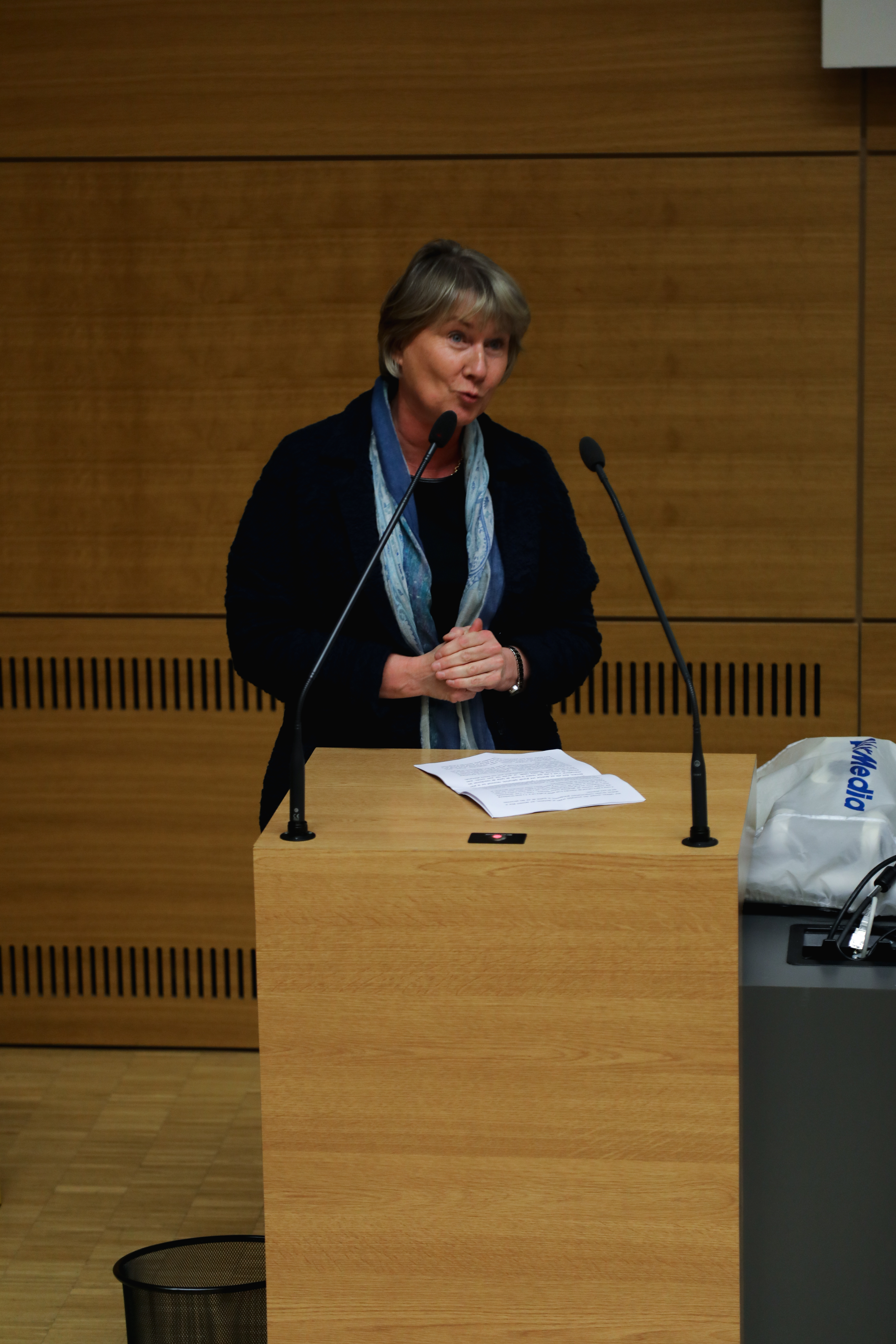
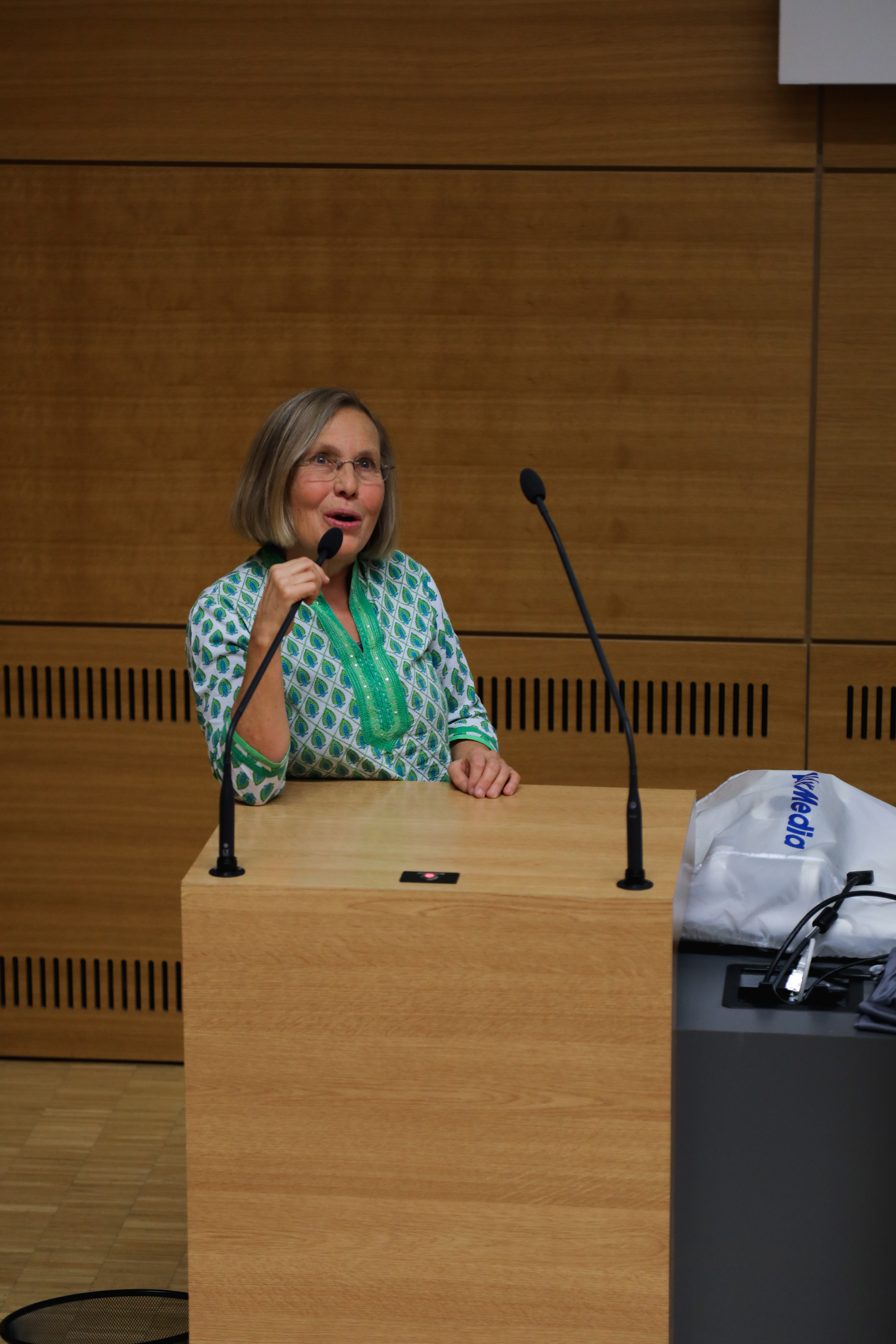
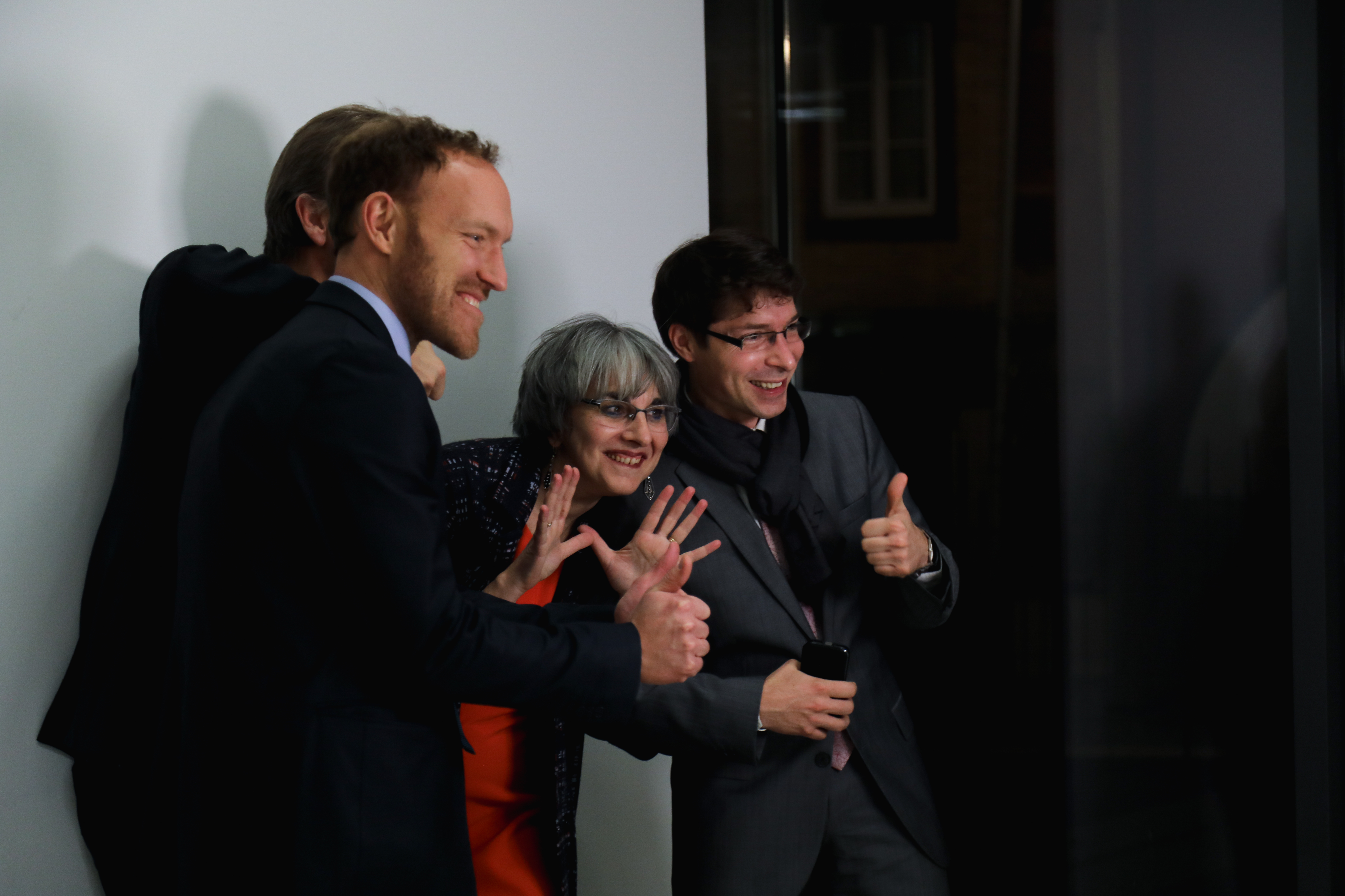
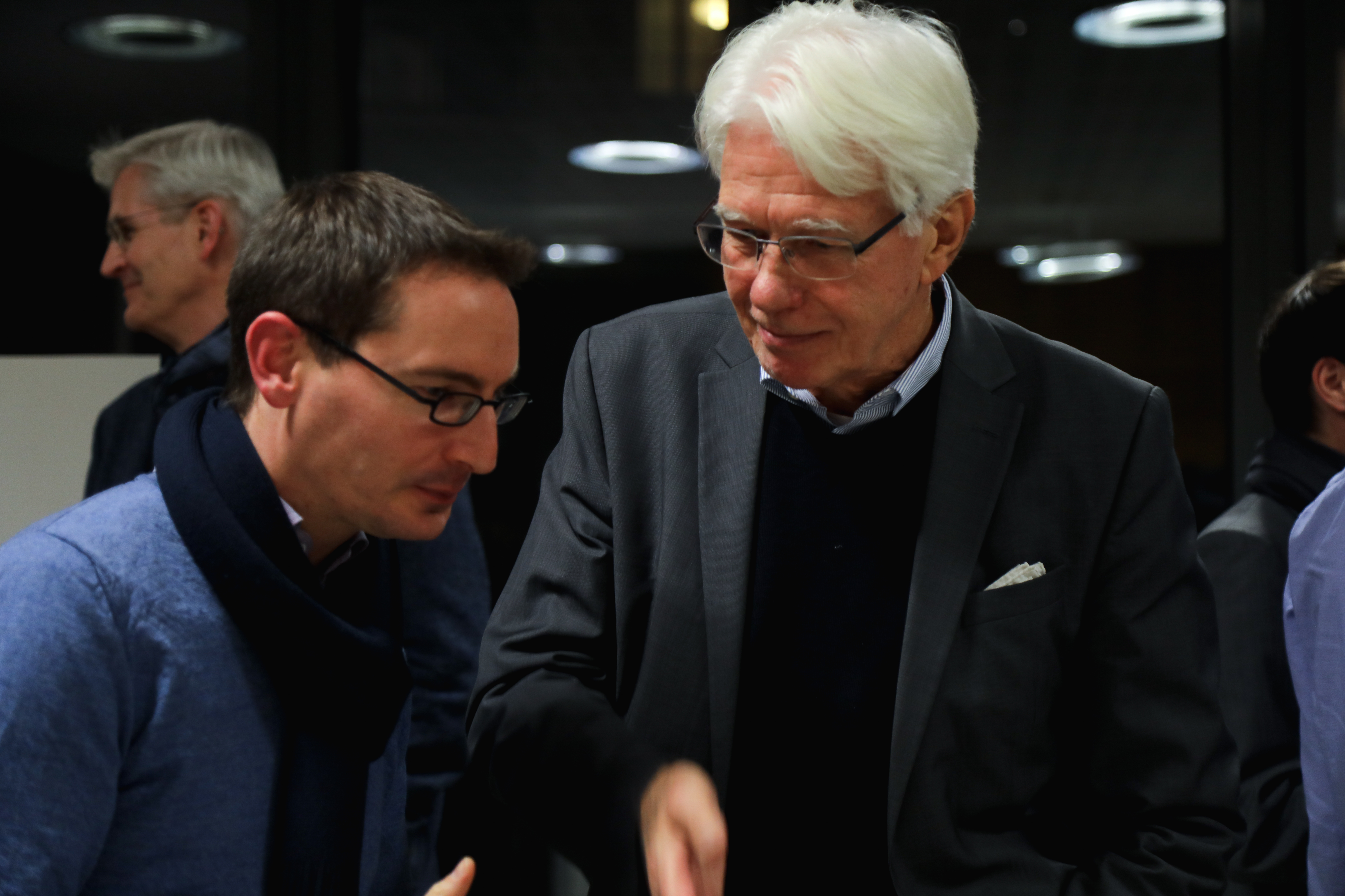
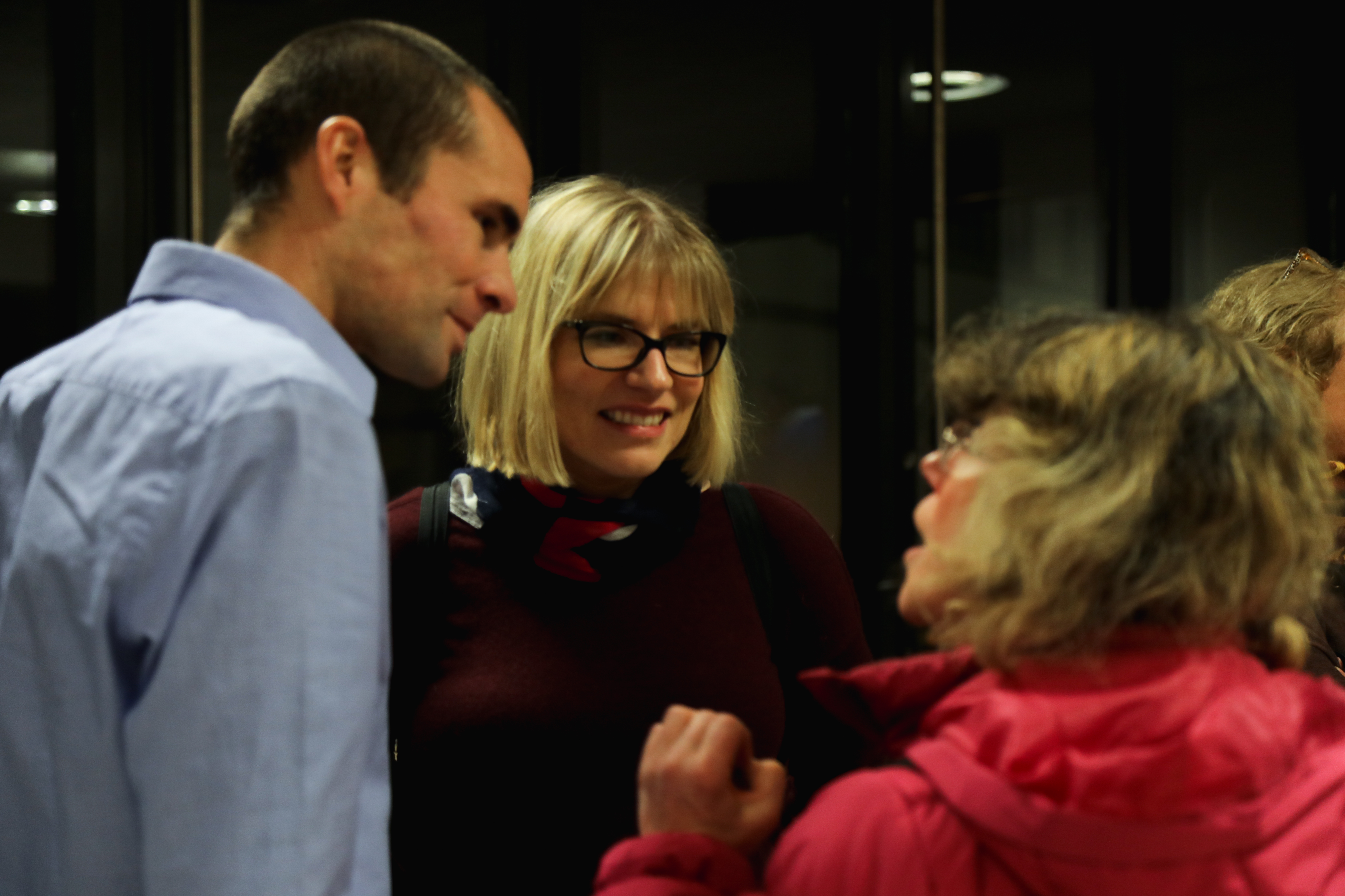
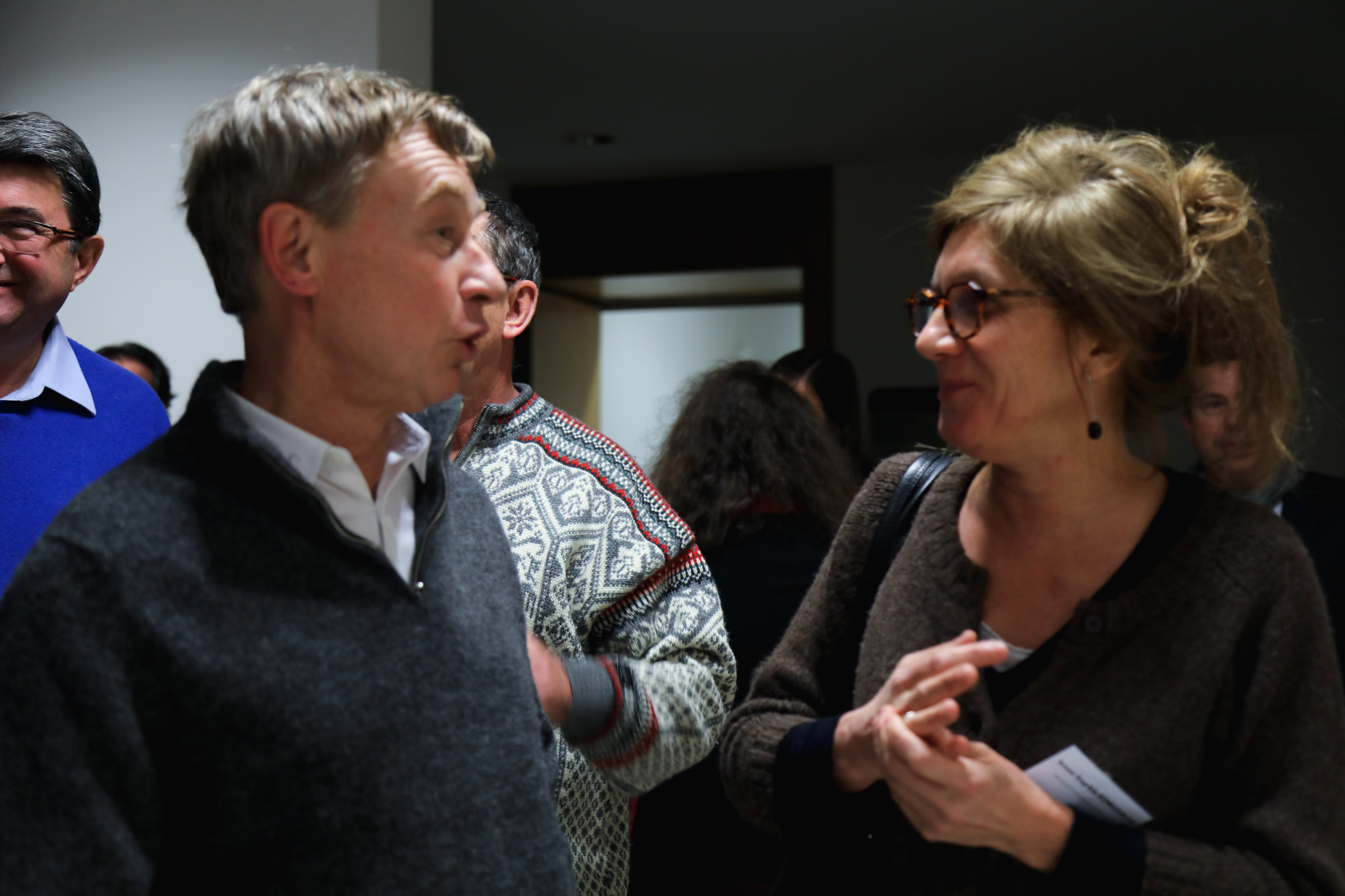
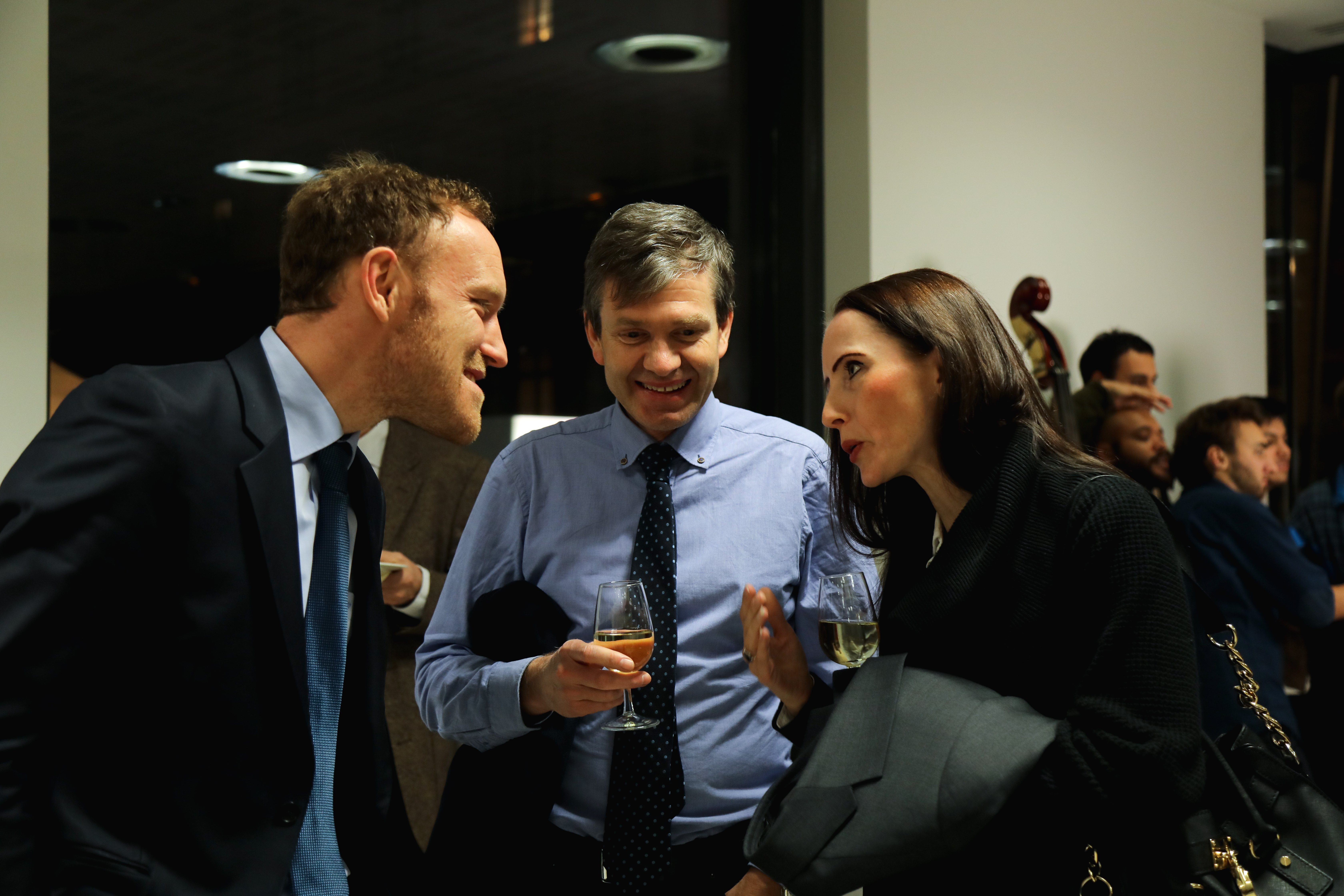
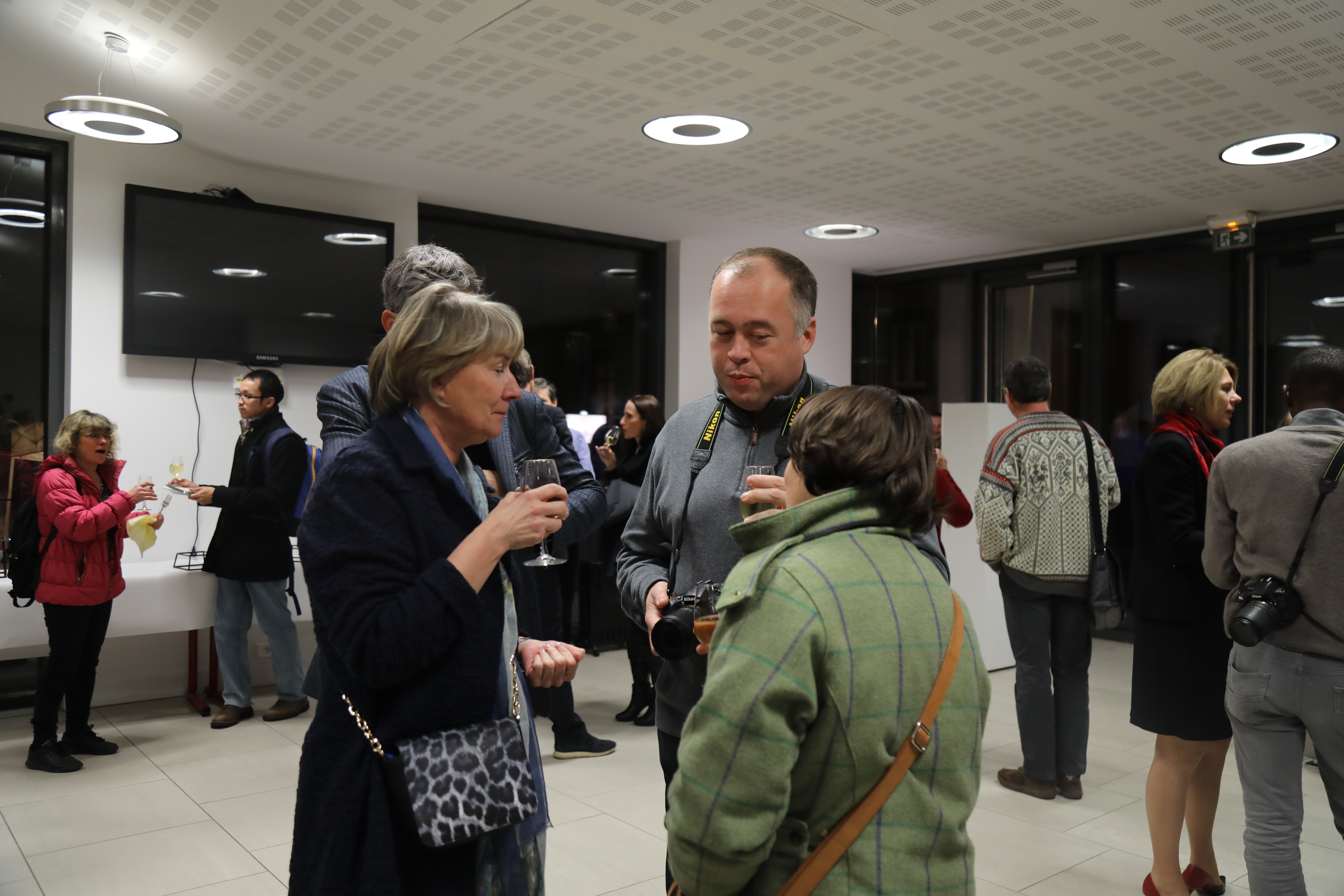
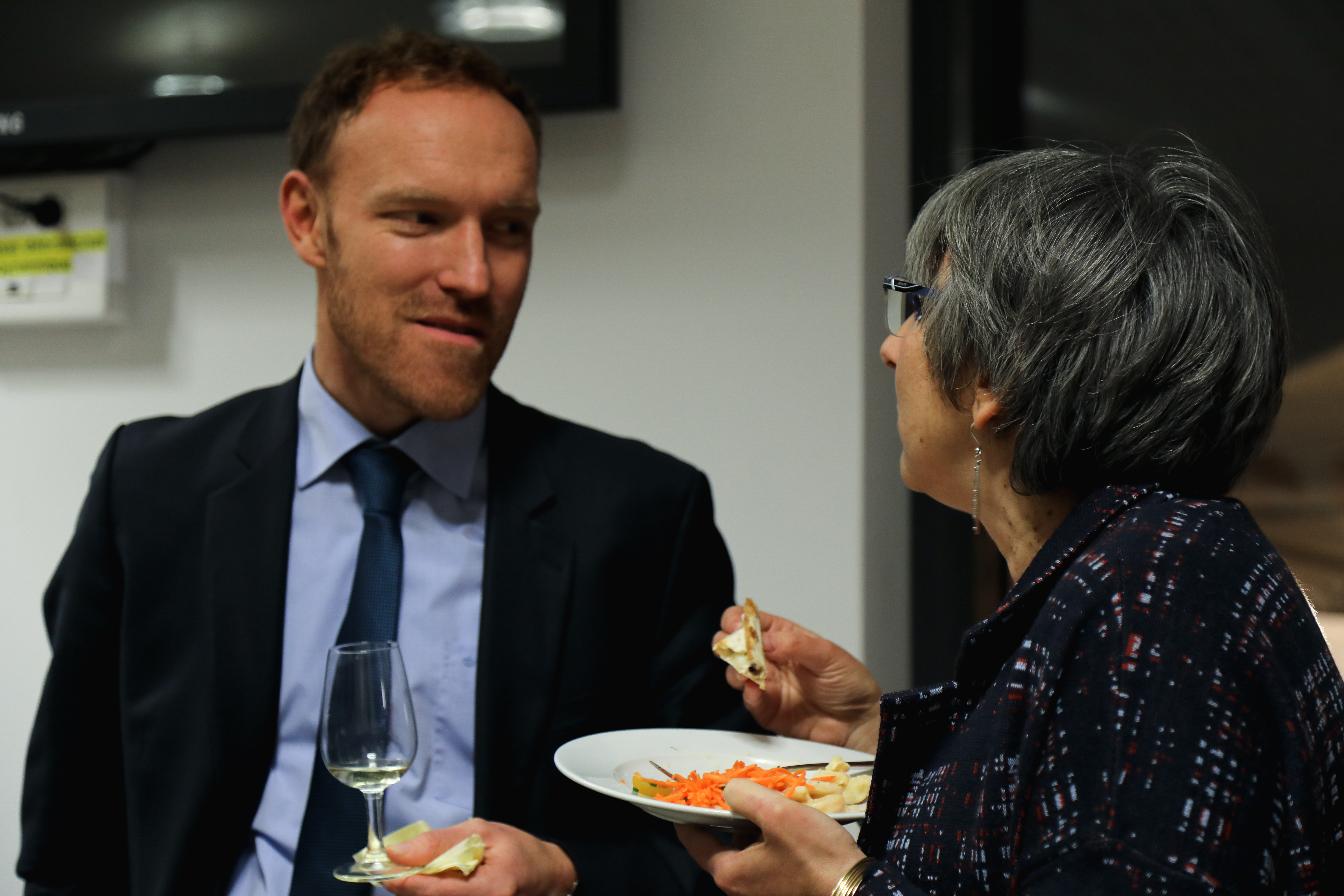
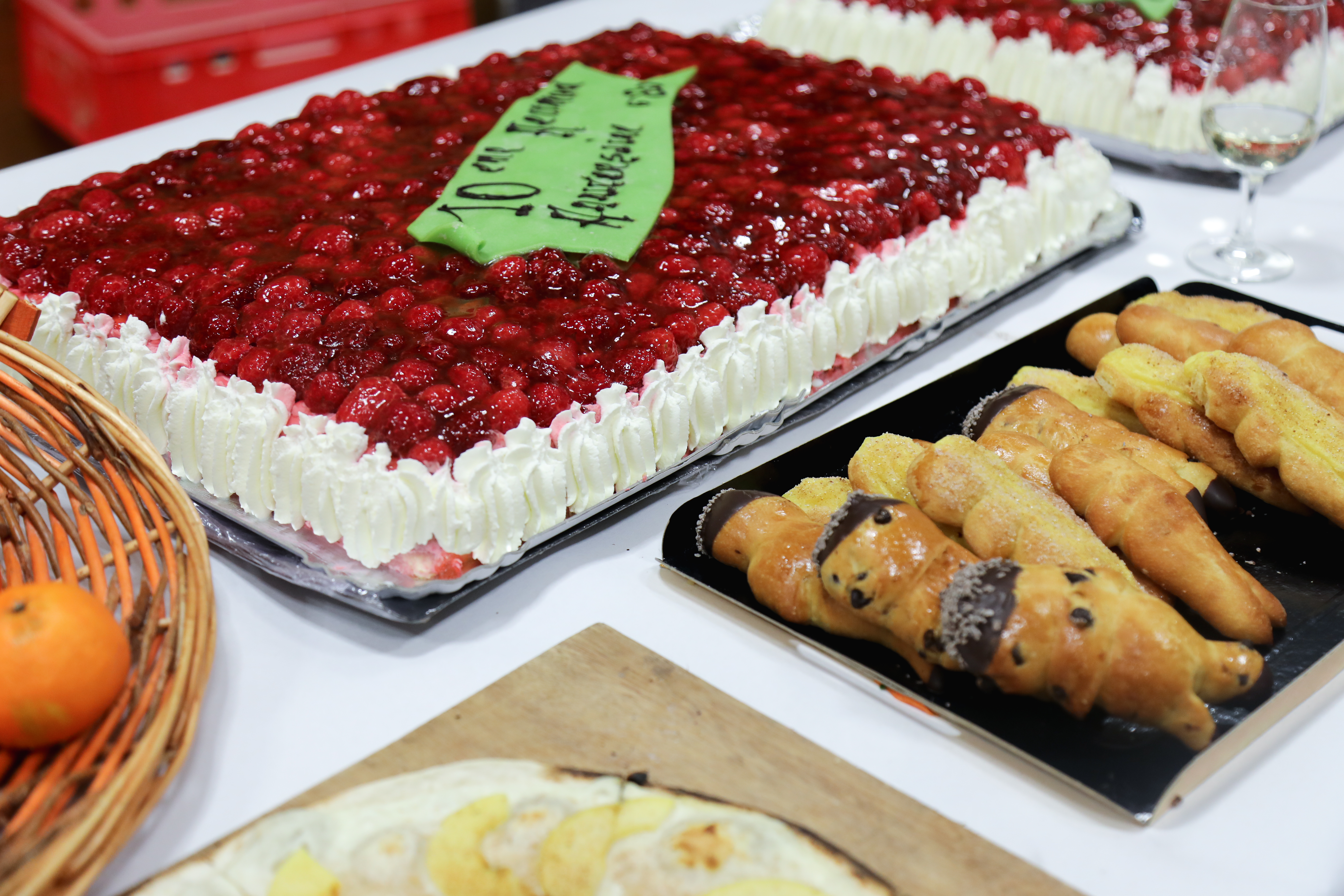
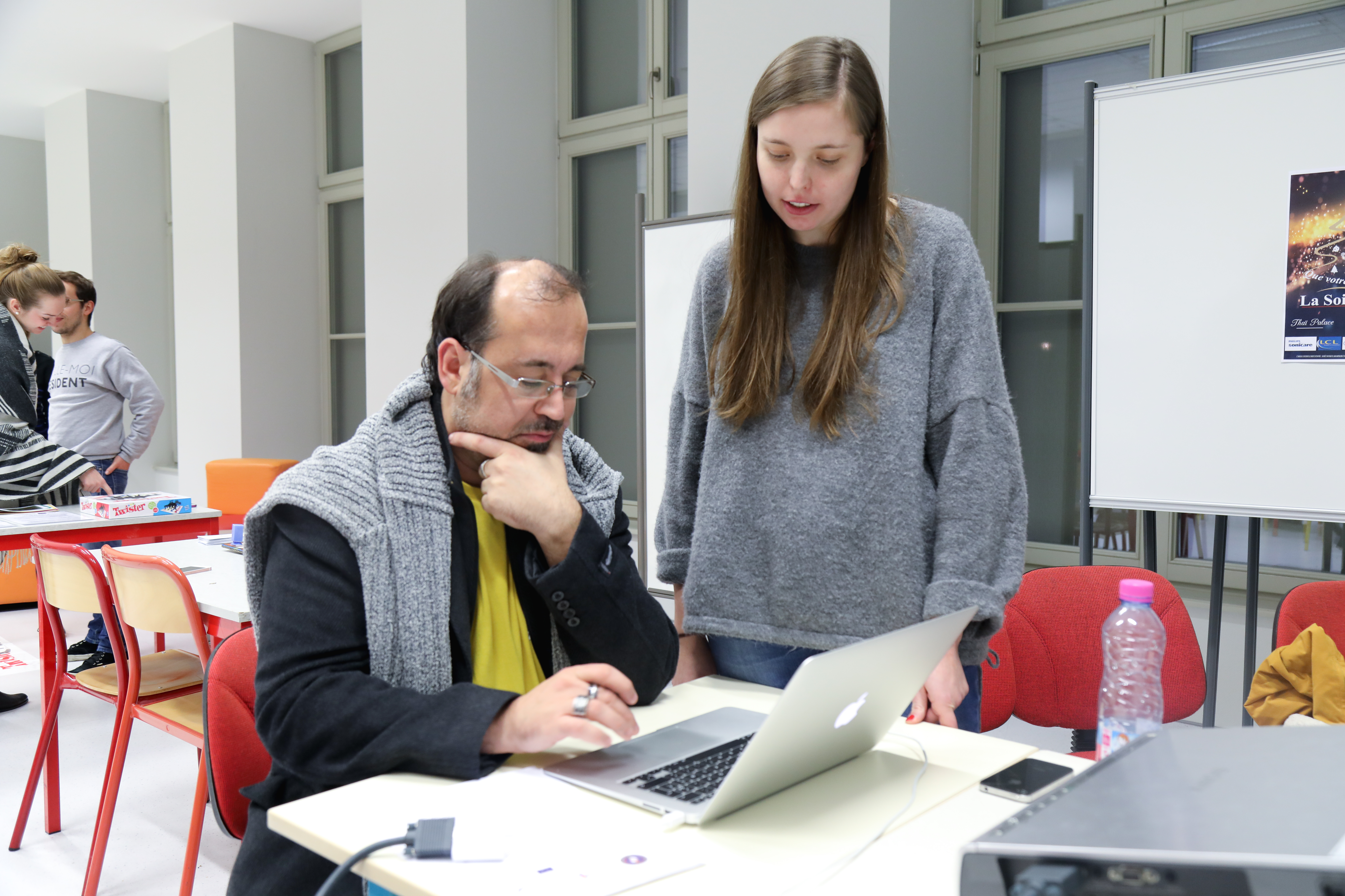
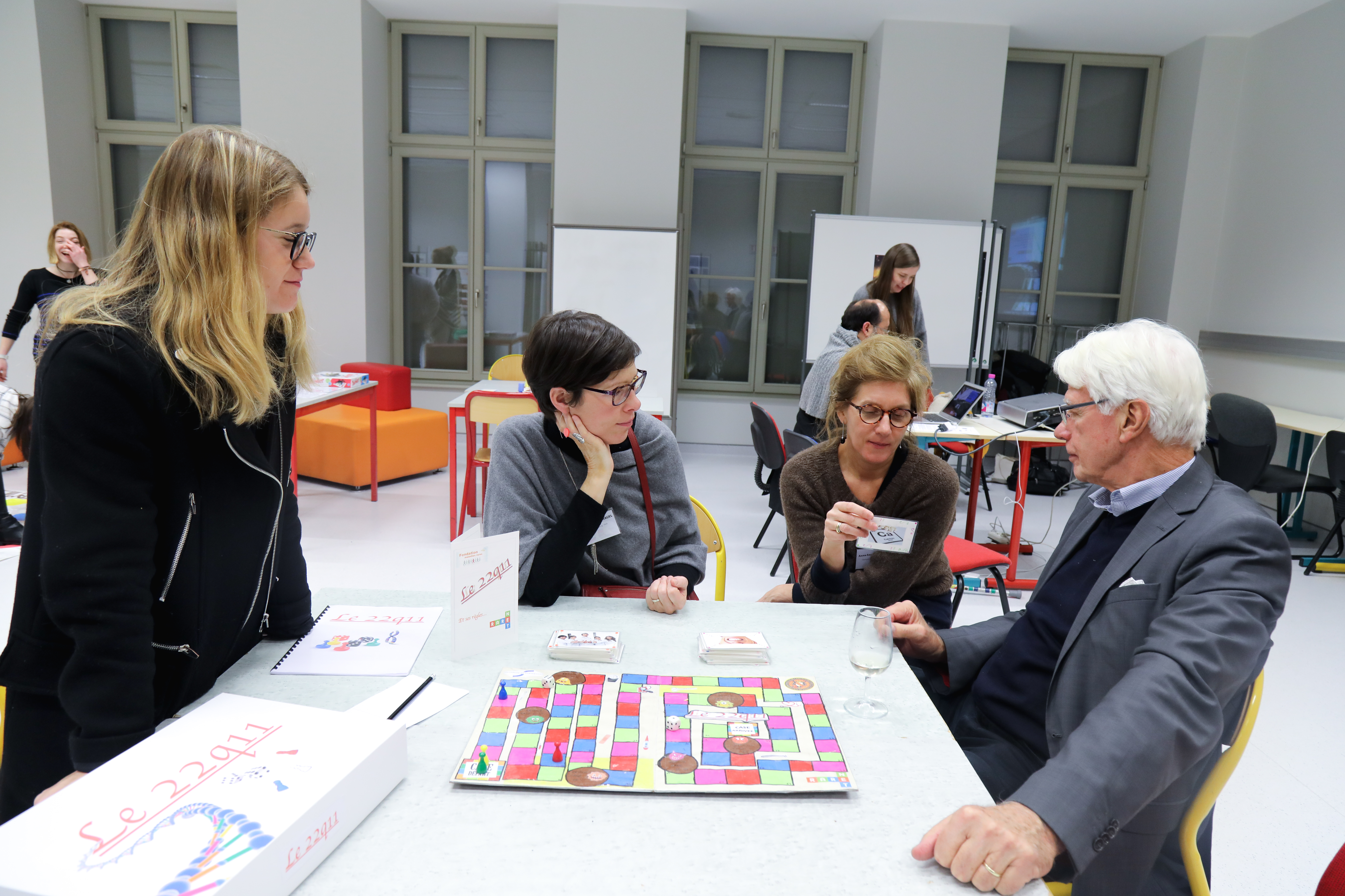
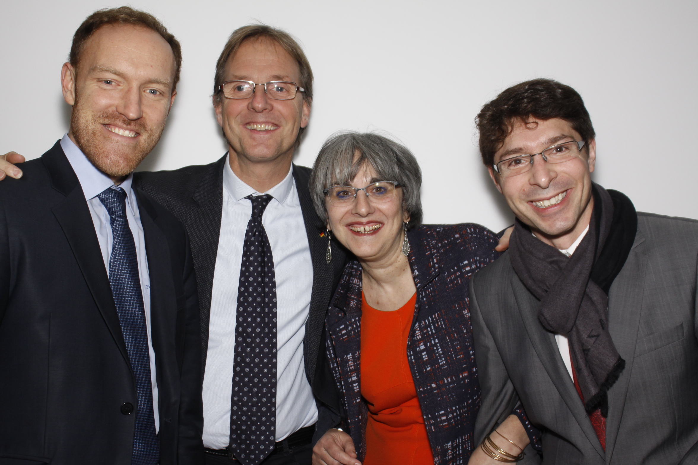
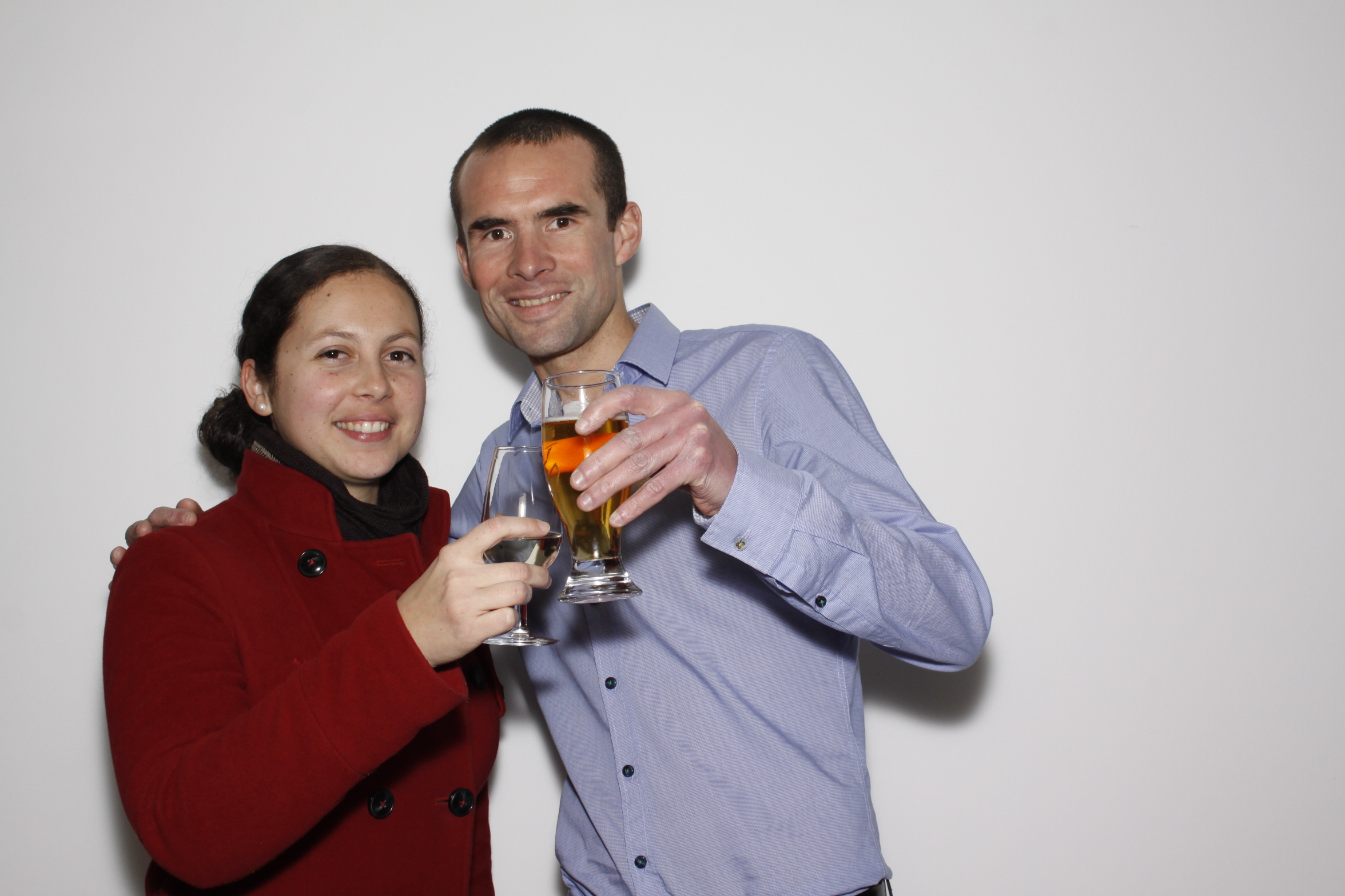
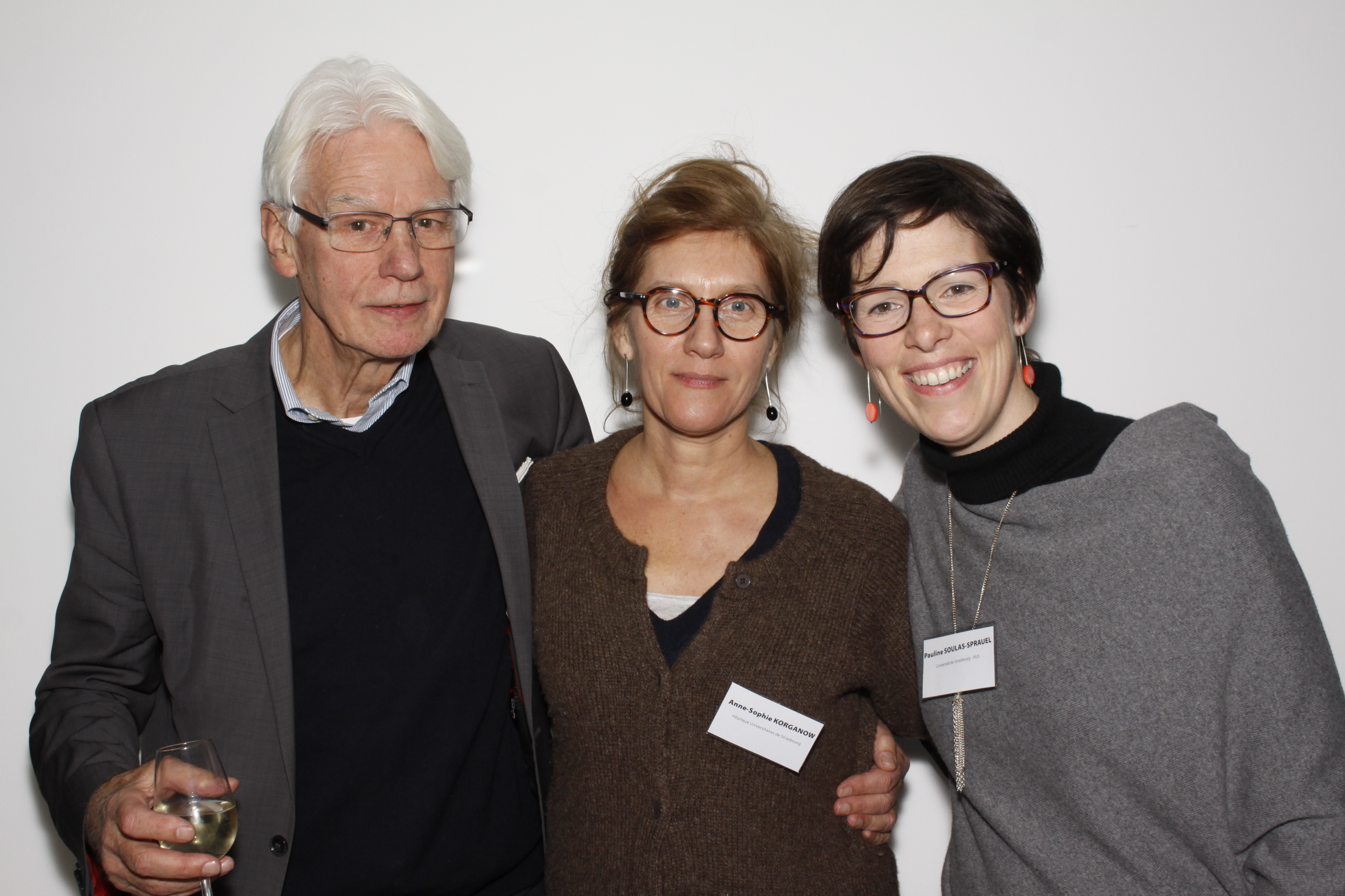
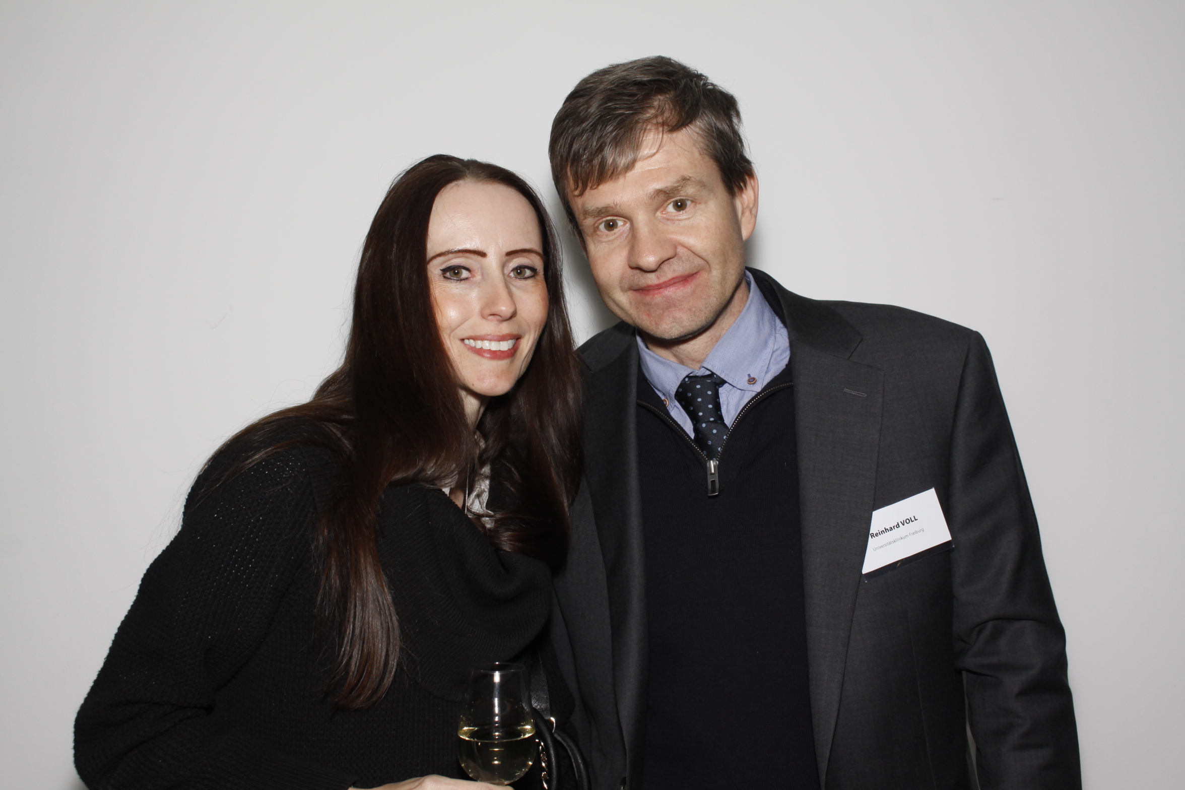
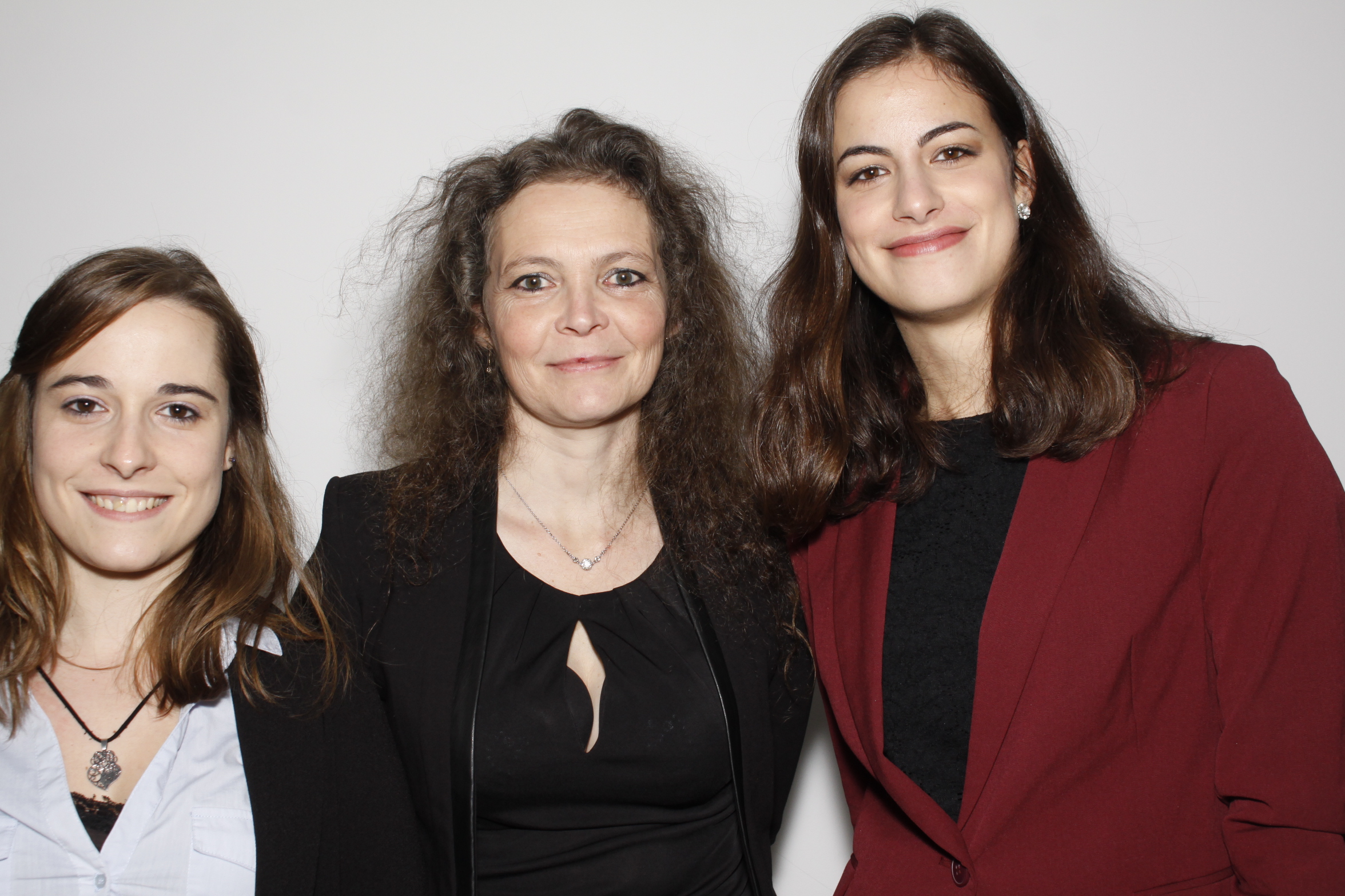
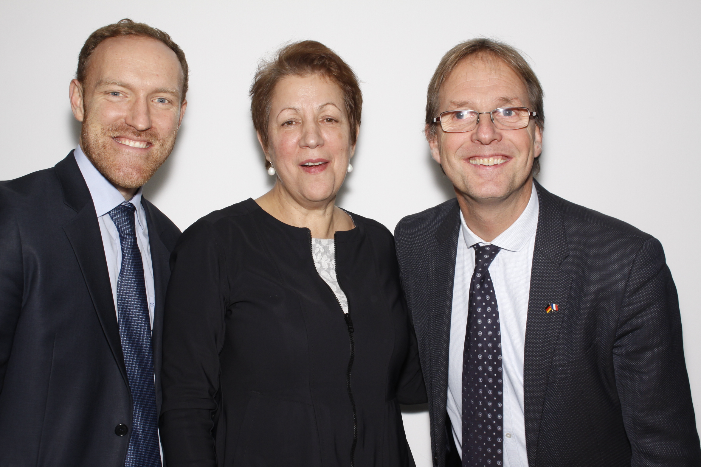
RARENET a célébré son lancement officiel les 7 et 8 décembre avec un symposium portant en parallèle sur les agénésies dentaires multiples et sur les maladies auto-immunes rares et vascularites. Une centaine de participants ont répondu à l’invitation ! Ce symposium très riche par la qualité des échanges et des contenus scientifiques ainsi que l’évènement festif ont été précédées d’une réunion de travail du consortium, le projet rassemblant une quarantaine de partenaires français, allemands et suisses.
Réunir des spécialistes des maladies rares auto-immunes et des manifestations bucco-dentaires des maladies rares et rassembler professionnels de santé, enseignants, chercheurs, industriels et associations de patients, telle est l’originalité du projet RARENET!
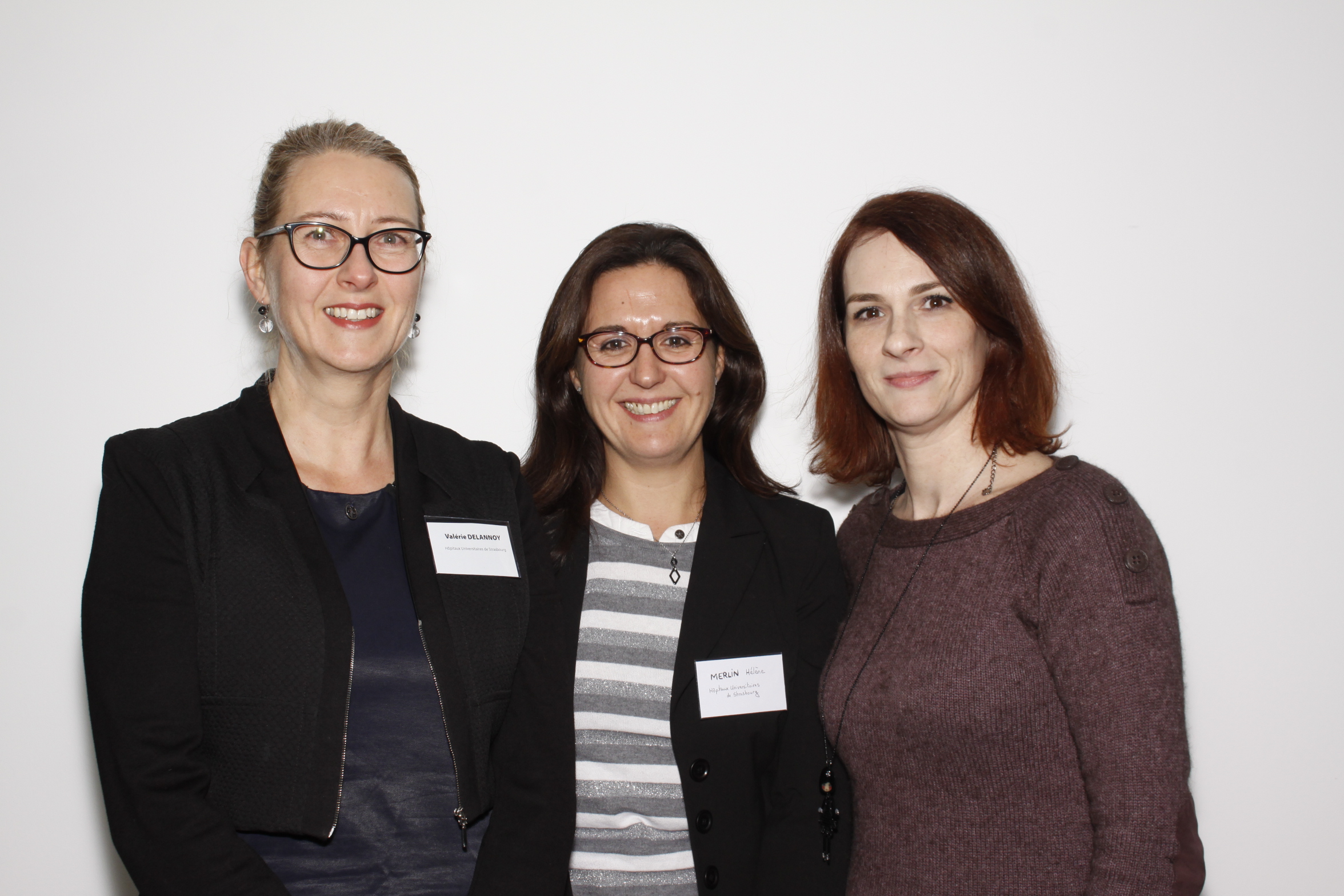
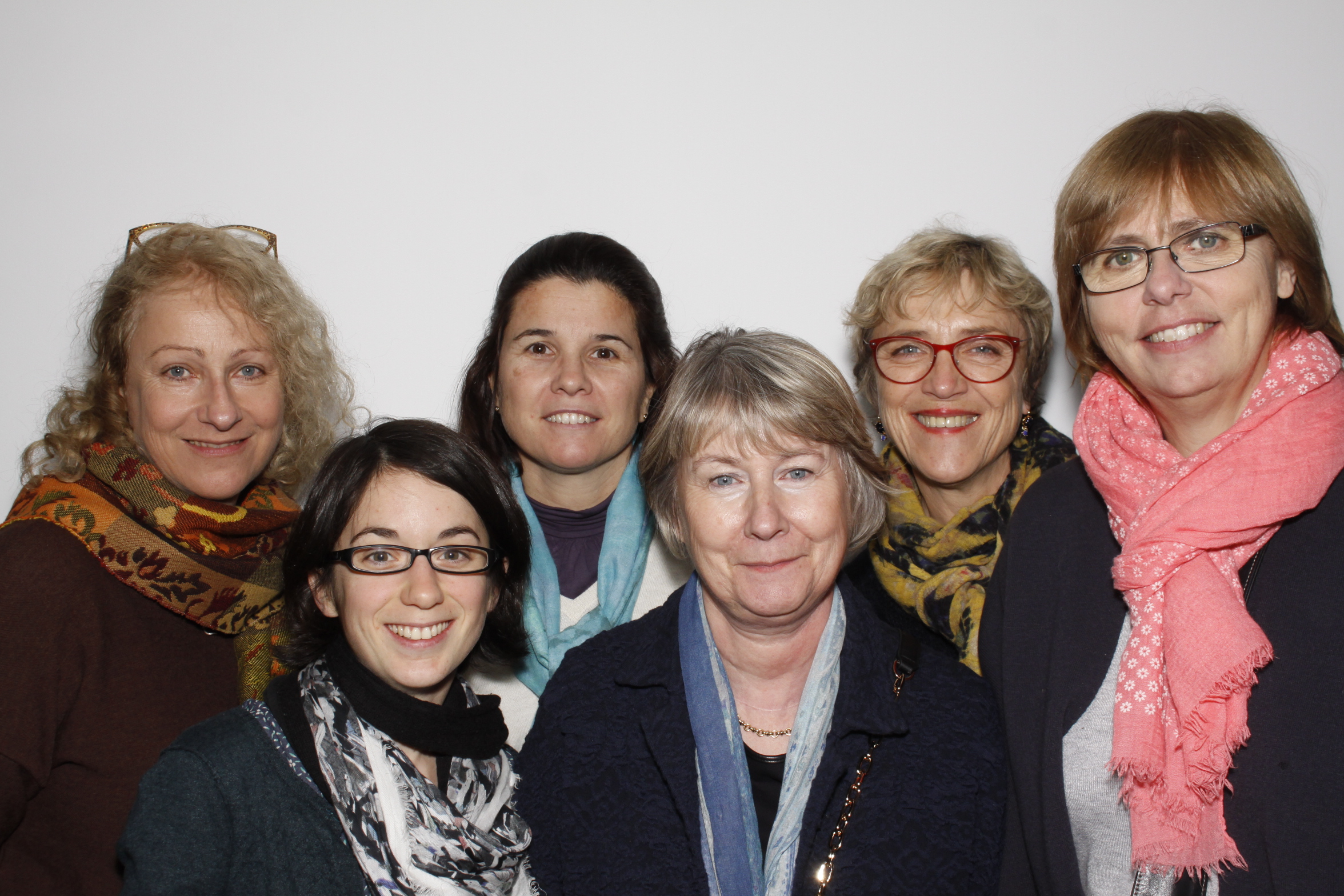
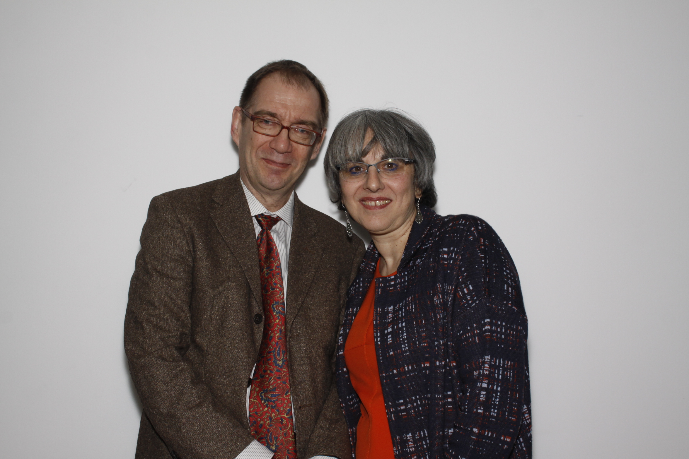
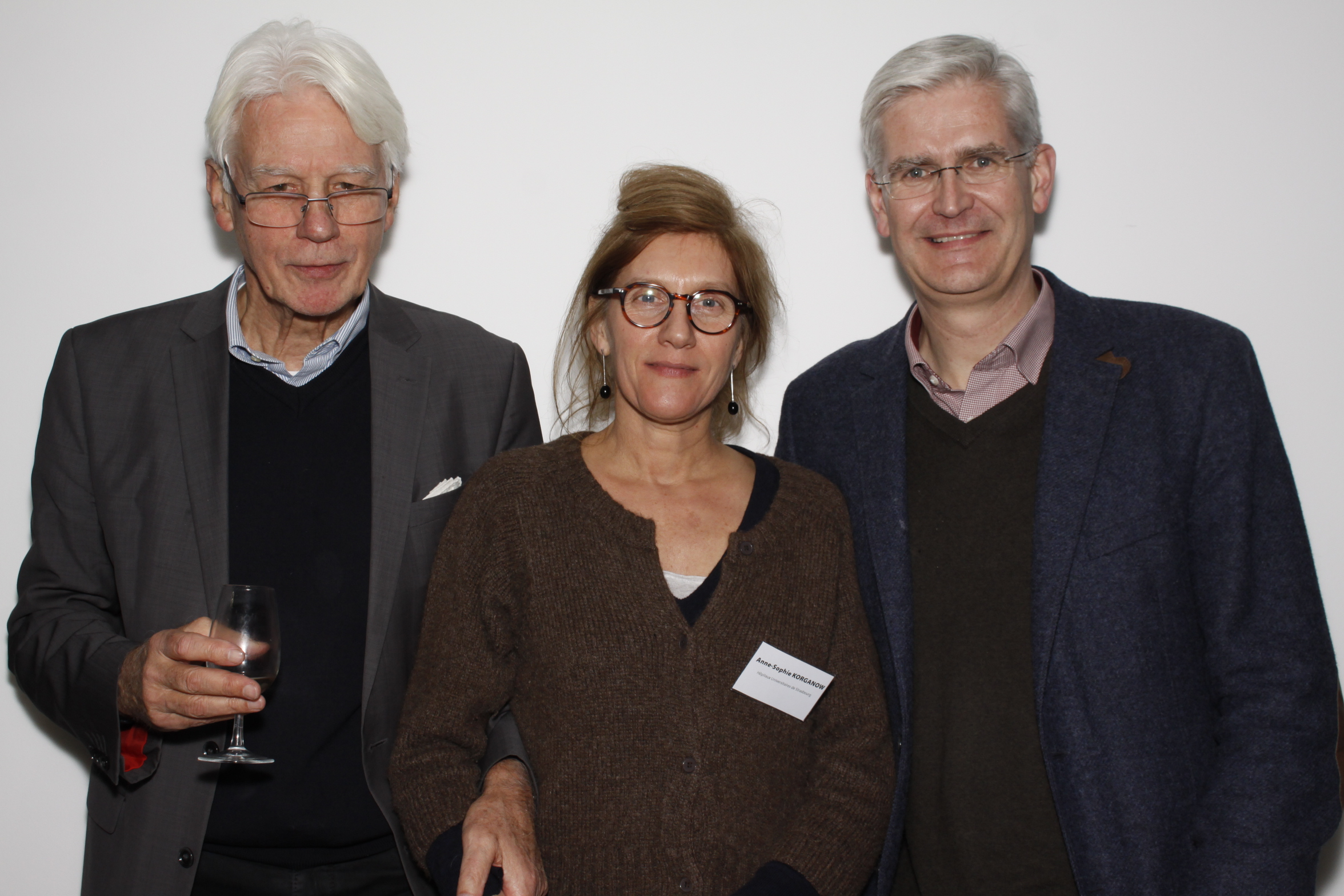
Voici quelques photos de cette belle soirée de lancement !

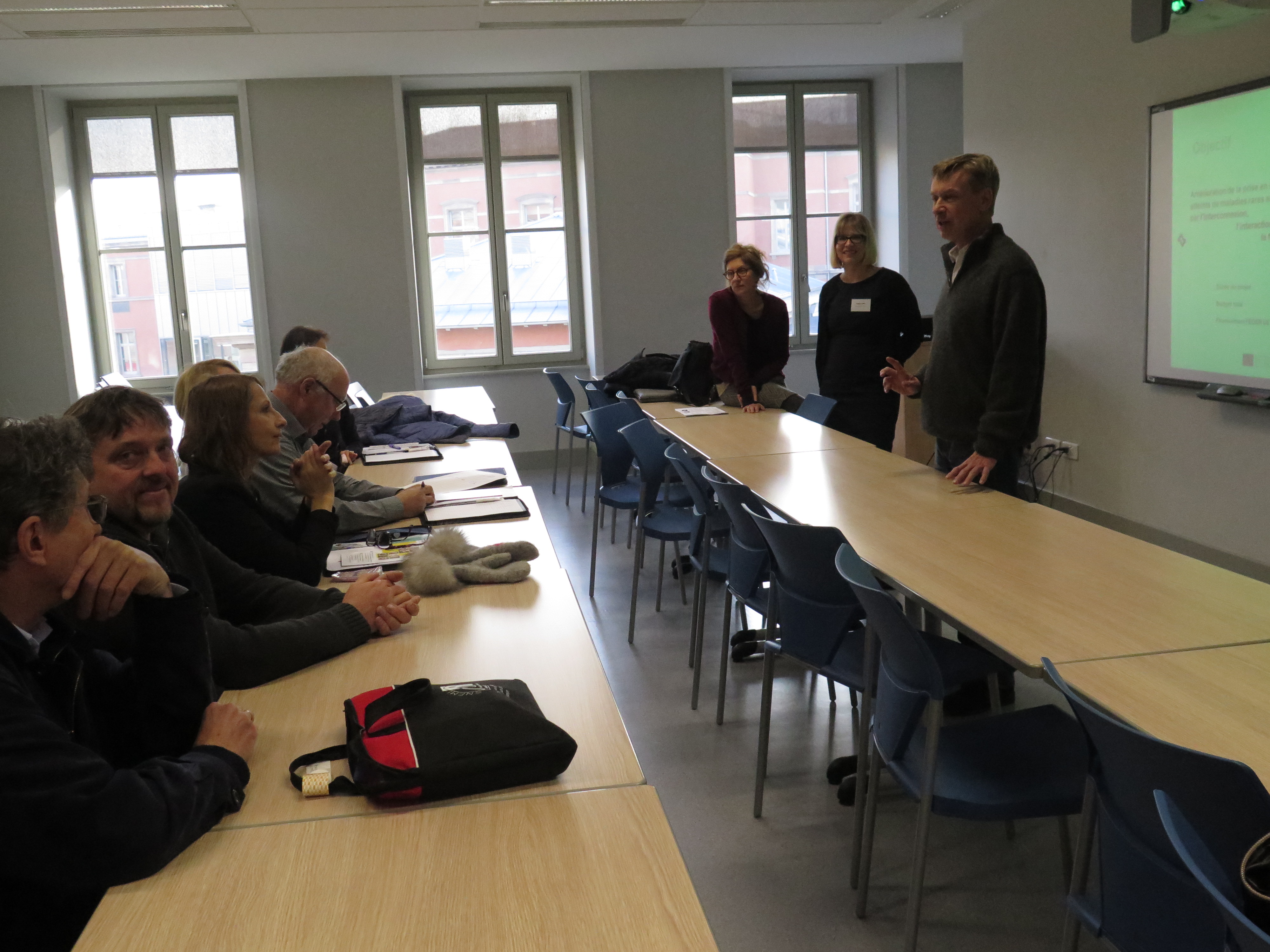
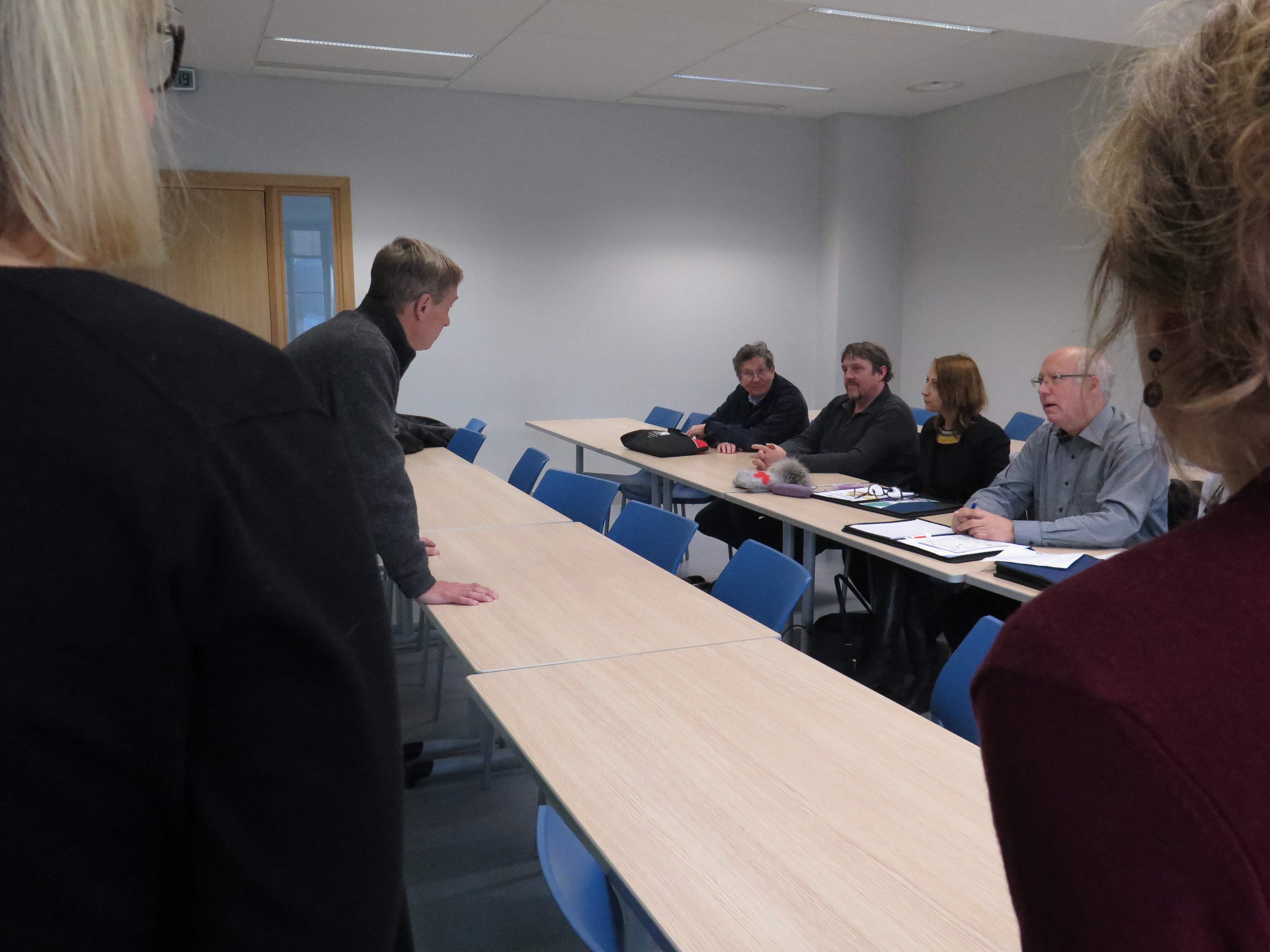
Les partenaires du projet se sont retrouvés pour leur deuxième réunion de consortium à Strasbourg le 7 décembre.
Ce moment d’échanges a permis de faire le point sur les questions de management et plus particulièrement sur la gestion financière et opérationnelle du projet.